Authors: Sebastian Seung
Connectome (8 page)

Lashley's doctrine has some element of truth but is too sweeping. The cortex is not infinitely adaptable. If it were, every stroke patient would recover completely. To understand the limits of adaptation and develop ways to enhance it, we need a deeper understanding. We know that the cortex can remap, but how exactly does the function of an area change?
We can't answer this without addressing a more basic issue: What defines the function of a cortical area in the first place? Broca's and Wernicke's regions are dedicated to language, and Brodmann areas 3 and 4 are dedicated to bodily sensation and movement. But
why
these functions? And how are they executed?
It's hopeless to answer these questions by studying only brain regions, their sizes, and their activity levels. We must look at the organization of the brain on a much finer scale. A cortical area can contain over 100 million
neurons. How are they organized to perform mental functions? In the next few chapters we'll explore this question, along with the idea that brain function depends heavily on the
connections
between neurons.
3. No Neuron Is an Island
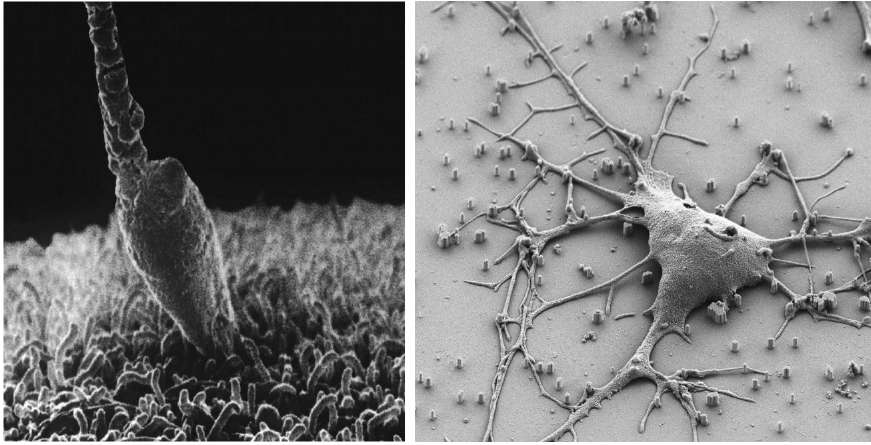
The neuron is my second-favorite cell. It's a close runner-up to my favorite: sperm. If you have never looked into a microscope to see sperm swimming furiously, grab your favorite biologist by the lapels of his or her lab coat and demand a viewing session. Gasp at the urgency of their mission. Mourn their imminent death. Marvel at life stripped down to its bare essentials. Like a traveler with a single small suitcase, a sperm carries little. There are mitochondria, the microscopic power plants that drive the whipping motion of its tail. And there is DNA, the molecule that carries the blueprint of life. No hair, no eyes, no heart, no brainânothing extraneous comes along for the ride. Just the information, please, written in DNA with the four-letter alphabet A, C, G, and T.
If your biologist friend is still game, ask to see a neuron. Sperm impress by their unceasing motion, but a neuron takes your breath away with its beautiful shape. Like a typical cell, a neuron has a boring round part, which contains its nucleus and DNA. But this
cell body
is only a small part of the picture. From it extend long, narrow branches
that fork over and over, much like a tree. Sperm are sleek and minimalist, but neurons are baroque and ornate (see Figure 13).
Â
 Â
Â
Â
Figure 13. My favorite cells: sperm fertilizing an egg
(left)
and a neuron
(right)
Â
Even in a crowd of 100 million, a sperm swims alone. At most one will achieve its mission of fertilizing the egg. The competition is winner take all. When one sperm succeeds, the egg changes its surface, creating a barrier that prevents other sperm from entering. Whether brought together by a happy marriage or a sordid affair, sperm and egg form a monogamous couple.
No neuron is an island. Neurons are polyamorous. Each embraces thousands of others as their branches entangle like spaghetti. Neurons form a tightly interconnected network.
The sperm and the neuron symbolize two great mysteries: life and intelligence. Biologists would like to know how the sperm's precious cargo of DNA encodes half the information required for a human being. Neuroscientists would like to know how a vast network of neurons can think, feel, remember, and perceiveâin short, how the brain generates the remarkable phenomena of the mind.
The body may be extraordinary, yet the brain reigns supreme in its mystery. The heart's pumping of blood and the lung's intake of air remind us of the plumbing in our houses. They may be complex, but they do not seem mysterious. Thoughts and emotions are different. Can we really understand them as the workings of the brain?
A journey of a thousand miles begins with a single step. To understand the brain, why not start with its cells? While a neuron may be a kind of cell, it is far more complex than any other. This is most obvious from its profuse branches. Even after many years of studying neurons, I am still thrilled by their majestic forms. I'm reminded of the mightiest tree on earth, the California redwood. Hiking in Muir Woods, or other redwood forests on the Pacific coast of North America, is a good way to feel small. You see trees that live for centuries or even millennia, enough time to grow to vertiginous heights.
Am I overreaching to compare a neuron to the towering redwood? In absolute size, yes, but consider further how these wonders of nature stack up against each other. The redwood's twigs are as thin as one millimeter, a width 100,000 times smaller than the tree's football-field height. A branch of a neuron, called a
neurite,
can extend from one side of the brain to the other, yet can also narrow to 0.1 micrometer in diameter. These dimensions differ by a factor of one
million.
In its relative proportions, a neuron puts a redwood to shame.
But why do neurons have neurites? And why do they branch to look like trees? In the case of a redwood, the reason for branches is obvious: The redwood's crown captures light, which is a source of energy. A passing sunbeam will almost surely collide with a leaf rather than travel all the way to the ground. Likewise, a neuron is shaped to capture contacts. If a neurite passes through the branches of another neuron, it will likely collide with one of them. Just as a redwood “wants” to be struck by light, a neuron “wants” to be touched by other neurons.
Â
Every time we shake hands, caress a baby, or make love, we may be reminded that human life depends on physical contact. But why do neurons touch? Suppose that the sight of a snake causes you to turn and run. You respond because your eyes are able to communicate a message to your legs:
Move!
That message is conveyed by neurons, but how?
Neurites are much more densely packed than the branches of a forest or even a tropical jungle. Think instead of a plate of spaghettiâor microscopically fine capellini. Neurites entangle much like the jumbled strands on your plate, allowing one neuron to touch many others. Where two neurons touch, there can be a structure called a
synapse,
a junction through which the neurons communicate.
But contact alone does not make a synapse, which most commonly transmits chemical messages. A molecule known as a
neurotransmitter
is secreted by the sending neuron and sensed by the receiving neuron. Secretion and sensing are performed by still other types of molecules. The presence of such molecular “machinery” signifies that a contact point is actually a synapse, as opposed to a place where one neurite just goes past another.
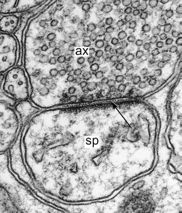
These telltale signs are blurred in an ordinary microscope, which uses light to make images, but show up nicely with a more advanced microscope based on electrons rather than light. The image shown in Figure 14 is a highly magnified (100,000Ã) view of a cut through brain tissue. There are two large, round cross-sections of neurites (marked “ax” and “sp”).
These are like the cut ends of strands that would be exposed if you sliced through spaghetti. The arrow points to a synapse between the neurites, which are separated by a narrow cleft. Now we see that the term
contact point
is not entirely accurate, as the neurites come extremely close to each other but do not really touch.
Â
 Â
Â
Â
Figure 14. A synapse in the cerebellum
Â
On either side of the cleft is the molecular machinery for sending and receiving messages. One side is dotted with many little circles, tiny bags called vesicles that store neurotransmitter molecules ready for use. On the other side the membrane holds a dark fuzz called the
postsynaptic density,
which contains molecules known as
receptors.
How does this machinery transmit a chemical message? The sender secretes by dumping the contents of one or more vesicles into the cleft. The neurotransmitter molecules spread out in the salty water there. They are sensed by the receiver when they encounter receptor molecules embedded in the postsynaptic density.
Many types of molecule are used as neurotransmitters. Each is assembled from atoms bonded to each other, as in the examples shown in Figure 15. (In these “ball-and-stick” models, each ball represents an atom and each stick a chemical bond.) You can see that each type of neurotransmitter has a characteristic shape determined by the specific arrangement of its atoms, a fact that will become important shortly.
Â
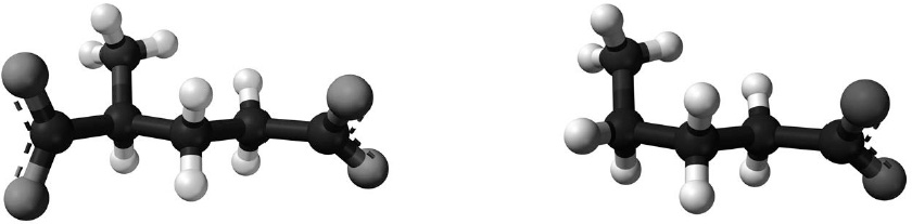 Â
Â
Â
Figure 15. “Ball-and-stick” models of neurotransmitters: glutamate
(left)
and GABA
(right)
Â
On the left is the most common one, glutamate. This is best known to the public in the form of monosodium glutamate (MSG), which is used as a flavor enhancer in Chinese and other Asian cuisines. Few realize that glutamate also plays a crucial role in brain function. Shown on the right is the second most common, gamma-aminobutyric acid, or GABA for short.
More than one hundred neurotransmitters have been discovered so far. The list sounds long. Do you ever feel overwhelmed in the liquor store, when you see the shelves stocked with so many brands of beer and wine? If you're a creature of habit, you might buy the same one or two brands every time and serve them to your friends at every party you give. That's what neurons do. With few exceptions, a neuron secretes the same small set of neurotransmitters
âoften only a single neurotransmitterâat all of its synapses. (The synapses in question are those made by a neuron onto others, not those received by a neuron.)
Now let's consider receptor molecules, which are much larger and more complex than neurotransmitters. Part of each molecule sticks out from the surface of the neuron, like the head and arms of a kid using an inner tube to float on water. This protrusion is the part of the receptor that senses neurotransmitter.
A glutamate receptor senses glutamate but ignores GABA and other neurotransmitters. Likewise, a GABA receptor senses GABA but ignores other molecules. Where does this specificity come from? Think of a receptor as a lock and the neurotransmitter as a key. As we saw above, each type of neurotransmitter has a distinctive molecular shape, which is like the pattern of bumps and grooves on a key. Every type of receptor has a location called the binding site, which has a characteristic shape like the innards of a hole in a lock. If the shape of the neurotransmitter matches that of the binding site, it activates the receptor, much as the right key in the right lock opens a door.
Once you know that the brain uses chemical signals, it's no longer surprising that drugs can alter the mind. A drug is a molecule too, and can be shaped like a neurotransmitter. If the mimicry is faithful enough, the drug will activate receptors, much as a copy of a key can open the same lock as the original. Nicotine, the addictive chemical in cigarettes, activates receptors for the neurotransmitter called acetylcholine. Other drugs inactivate receptors, much as an inaccurate copy of a key might turn partially and jam the lock. Phencyclidine or PCP, known on the street as “angel dust” in honor of its recreational use for hallucinogenic effects, inactivates glutamate receptors.