The Cerebellum: Brain for an Implicit Self (63 page)
Read The Cerebellum: Brain for an Implicit Self Online
Authors: Masao Ito
Tags: #Science, #Life Sciences, #Medical, #Biology, #Neurology, #Neuroscience

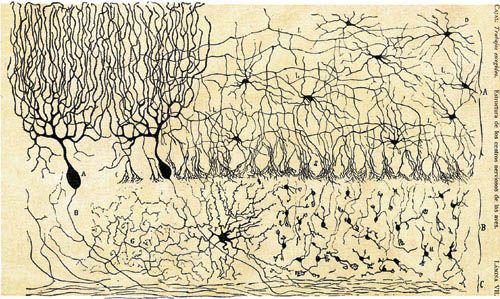
Color Plate IV. A classic Ramón y Cajal drawing of cerebellar cells.
Shown are Purkinje (A, B), stellate (D), Golgi (F), and granule (H) cells in the cerebellum. Also note the basket-cell axons (S) terminating freely around the Purkinje cell bodies. (Courtesy of the Instituto de Neurobiolog’a “Ramón y Cajal,” Madrid, Spain.)
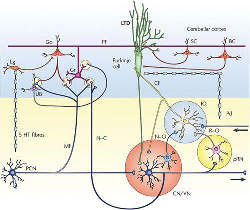
Color Plate V. Sketch of neuronal circuits in the cerebellum.
Long-term depression of Purkinje cells is induced by the conjunctive activation of parallel fibers and climbing fibers. (From
Ito, 2009
.)
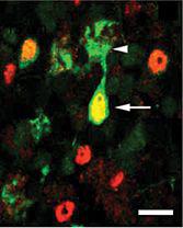
Color Plate VI. Unipolar brush cells in the adult mouse cerebellum.
Shown are orange-stained cell bodies of unipolar brush cells, using the transcription factor, Tbr2/E, as the marker. Also shown is one unipolar brush cell of the calretinin subtype, which is green-stained in the outer edges of the cell body (at arrow) and in the dendritic brush (arrowhead) with an anticalretinin antibody. The yellow staining in the center of the cell body is due to staining with Tbr2+. Calibration bar, 13 micrometers. (From
Englund et al., 2006
.)
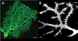
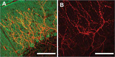
Color Plate VII. Purkinje cell dendrites.
Shown are the dendrites of two rat Purkinje cells injected with Alexa-594 from another rat. (A) A Purkinje cell on Z-stack image obtained from optical sections using a confocal laser scanning microscope. Scale, 20 micrometers. (B) Purkinje cell dendrites reconstructed three-dimensionally. Scale, 2 micrometers. (Courtesy of Tetsuya Tatsukawa.)
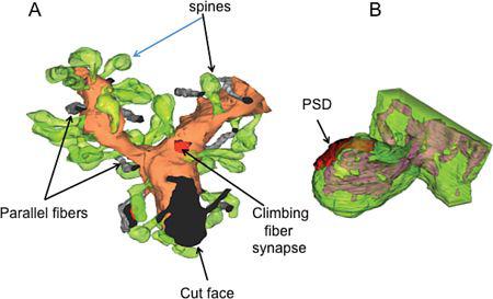
Color Plate VIII. Dendritic spines of Purkinje cells.
(A) A dendrite segment from an adult rat Purkinje cell, reconstructed from consecutive 90 nm serial sections. In this 3D rendition, the dendritic shaft (orange) at a branch point is studded with synaptic spines (in semitransparent green). Only a subset of the parallel fibers (gray) crossing the dendritic plane is shown here, with one displaying the typical “en passant” synaptic contact, with its postsynaptic density (PSD) shown in red (lower-left quadrant). In this reconstruction, only two climbing fiber synapses were found, with one visible here, as identified by its PSD (also red) in apposition with the Purkinje cell dendritic shaft. (B) A single spine reconstructed by electron tomography. The complex branched structure of the spine’s endoplasmic reticulum (purple) is visible through the semitransparent representation of the plasma membrane. In the dendritic shaft, note the continuity with endoplasmic reticulum. In A, average spine length is 1.36 micrometers. The spine in B is 1.4 micrometers long—i.e., from the base of its neck to its apex. (Courtesy of Thomas Launey.)
Color Plate IX. Climbing fibers.
After the injection of the tracer, BDA (biotinylated dextran amine), into the inferior olive of a mouse, labeled climbing fibers are visualized with immunofluorescent (red) methods in the cerebellar cortex. (A) Purkinje cells were also visualized by immunohistochemistry for a calcium binding protein, Calbindin (green). (B) Without labeling Calbindin. Scale bars: 100 micrometers. (Courtesy of Tsutomu Hashikawa.)
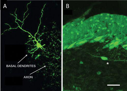
Color Plate X. A Golgi cell and a Lugaro cell.
(A) This Golgi cell in a cerebellar slice was filled with AlexaFluo through a patch-clamp pipette (removed). It was reconstructed into a stack view by use of a confocal microscope. Note the cell’s broad extension of its axonal plexus, multiple short basal dendrites, and two apical dendrites, which climb into the molecular layer (not shown). (B) * marks a Lugaro cell observed under a microscope in a cerebellar slice derived from a gene-manipulated mouse that expressed GFP specifically in GABAergic neurons. Note also the green-labeled Purkinje and stellate cells, including their dendrites, in the molecular and Purkinje cell layers. Scale for B, 50 micrometers. (A, from
D’Angelo, 2008
; B, courtesy of Moritoshi Hirono.)
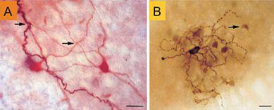
Color Plate XI. Gap junction couplings among inferior olive neurons.
(A) Dye injection into a “straight” (term based on shape of dendritic morphology) neuron resulted in indirect labeling of nine additional neurons, two of which (also straight) are shown in this panel. The darkly stained dendrite belongs to the cell that was labeled directly. Arrows denote examples of intersections of the dendrites of different cells, these being the possible locations of gap junctions. (B) Dye injected into a curly neuron (note its large dark cell body) resulted in indirect labeling of curly neurons. Indirectly labeled neurons had clearly stained cell bodies (e.g., the one at the arrow), but very weakly labeled dendrites. Scale bars, 20 micrometers. (From
Devor and Yarom, 2002
.)
Color Plate XII. A Ca
2+
surge induced by conjunctive stimulation in a Purkinje cell.
(A) This cell was visualized using infrared differential interference contrast optics. A patch pipette containing a Ca
2+
indicator was attached to the cell’s soma. Parallel fibers (PF) were stimulated using an extracellular electrode placed in the molecular layer. The dendritic region enclosed in the red-line rectangle was receiving synapses from the stimulated PFs as indicated by an increase in the fluorescent signals. Conjunctive stimulation was performed with a combination of 2 PF-stimulation at 30 ms intervals and somatic depolarization from– 70 to –20 mV for 150 ms, repeated at 1 Hz for 5 minutes. (B) Ca
2+
images in the red-line rectangle in A shown before (a) and then 0.5, 2.5, and 4.5 (b, c, and d) minutes after the onset of conjunctive stimulation. (C) Changes in fluorescence intensity as a function of time, these being normalized relative to the average intensity recorded before conjunctive stimulation. Curves recorded for 10 seconds at times a–d in B are shown in different colors (a in black, b in red, c in green, and d in brown). Note that the curves in c and d nearly overlap each other. (Supporting data for
Le et al., 2010
.)