Read Equine Massage: A Practical Guide Online
Authors: Jean-Pierre Hourdebaigt
Equine Massage: A Practical Guide (9 page)

(34,
(35,
(36,
(37) superficial digital flexor tendon
(38) ob
(39) medial car
(40) mid
les
il wing
usc
le
usc
le
le
le
nocephalic
le
usc
elid
yoid m
usc
usc
usc
le
e
le
le and tendon
rate m
le
usc
usc
le
usc
t of ster
les of the Horse
al ser
usc
usc
al nasal m
entr
apezius m
apezius m
le of upper lip and its tendon
le
le
le of lower ey
le of the ey
les
le of the mouth
le of lower lip
le
ular par
le
usc
yoid and omoh
le of upper lip and nostr
il dilator m
usc
usc
usc
usc
usc
t of rhomboid m
t of v
t of tr
t of tr
usc
usc
usc
usc
hiocephalic m
apezius m
usc
t of later
yroh
ac
ator m
erse nasal m
le and tendon
al nostr
rugator supercilii m
icular m
noth
nomandib
acic par
vical par
vical par
vical par
ts of tr
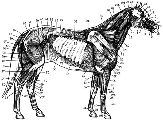
Superficial Musc
ator m
ansv
usc
uccinator m
m
17a) br
par
1a) lev
1.13
(1,
(2) lev
(3) dorsal par
(4) later
(5) orbicular m
(6) b
(7) depressor m
(8) zygomatic m
(9) masseter m
(10) tr
(11) depressor m
(12) orbicular m
(13) cor
(14) aur
(15) ster
(16) ster
(17,
(18) cer
(19) splenius m
(20) cer
(21) cer
(22) thor
(22a) tendinous intersection between the two
30
Equine Massage
m
anial
tion of the cr
les
les
usc
usc
les
usc
ator m
le (cunean tendon or cuneifor
ator m
usc
tion)
t tail lev
tibial m
inser
64a) annular ligaments
(63a) medial tendon of inser
(64,
(65) shor
(66) long tail lev
(67) tail depressor m
les)
usculus
usc
t of
ior par
usculus semitendi-
ing group of m
le (anter
usc
is and m
y or tarsal tendon (from m
anial tibial m
biceps femor
nosus of the hamstr
flexor metatarsi)
(62) accessor
(63) cr
.,
(continued)
i.e
les)
usc
les of the Horse
y or tarsal tendon
hed to tuber calcanei,
superficial digital flexor
ing m
le
le
usc
usc
and accessor
hilles tendon,
Superficial Musc
of tendons attac
Ac
tendon,
from the hamstr
1.13
(59) soleus m
(60) popliteal m
(61) common calcaneal tendon (the aggregate
Anatomy and Physiology of the Horse
31
Skeletal muscles are highly elastic and have strong contractile power.They respond to motor nerve impulses; as a result, the contraction mechanism is a generated process. The release process is not a generated process, but rather is a natural relaxation of the muscle as a result of the cessation of the motor nerve impulses that originally “asked” the muscle to contract.
When a muscle develops a
contracture,
the muscle fibers stay contracted, eventually resulting in a spasm—at which point the natural relaxation process will not happen. Pain and motion problems will develop as a result.
Muscles are equipped with two types of sensory nerve endings: the Golgi apparatus and the muscle spindle. The
Golgi apparatus
nerve endings send feedback impulses to the brain as to the whereabouts of the muscles; this process is referred to as
proprioception.
The Golgi nerve endings are mostly located where the muscles and the tendons come together.
The nerve endings of the muscle spindle prevent overstretching of the muscle fibers. As its name implies, this nerve fiber coils around the length of the muscle bundle. Reaching a given length, the muscle spindle fires nerve impulses that trigger a fast reflex motor nerve reaction to induce immediate contraction of the muscle fibers. Thus the overstretching and potential tearing of fibers is prevented.This is a safety reflex mechanism.
When a muscle overstretches, a spasm often results. This is a
tetanic
(violent) contraction of a muscle in response to overstretching or trauma, whereby the muscle is unable to release its rigidity. A
microspasm,
on the other hand, is a small spasm occurring in just a few fibers of the muscle bundle. Microspasms have a cumulative effect over a period of time, resulting in a full spasm.
Sometimes a muscle is stretched past its limit and muscle fibers will tear. This causes an immediate muscle spasm and triggers an inflammation response, with swelling at the site of injury. As part of the healing process, new connective tissue is laid down in an irregular, scattered pattern within the muscle fiber arrangements.
Unfortunately this scar tissue reduces the muscle’s tensile strength, flexibility, and elasticity. Massage therapy can reduce the amount of scar tissue by applying deep kneadings and frictions after proper warm-up of the tissues. Also, stretching is a great technique for prevention and reduction of the formation of scar tissue.
A heavily exercised muscle will often develop light inflammation within its fibers. This is a normal process that promotes formation of new muscle fibers. But it is important to keep any inflammation under control to avoid the formation of scar tissue.
To keep inflammation down, use cold hydrotherapy, ultrasound,
32
Equine Massage
Anatomy and Physiology of the Horse
33
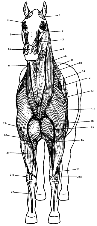
1.14 Muscles of the Horse, Front View
(1, 1a) levator muscle of upper lip and tendon
(2) levator muscle of upper lip and nostril wing
(3) lateral nostril dilator muscle
(4) corrugator supercilii muscle
(5) interscutular muscle
(6) brachiocephalic muscle
(7) sternomandibular part of sternocephalic muscle
(8) omohyoid muscle
(9) sternothyroid muscle
(10) cervical part of trapezius muscle
(11) jugular vein
(12) supraspinous muscle
(13) remains of skin muscle in the neck
(14) cranial deep pectoral muscle
(15) cranial superficial pectoral muscle
(16) caudal superficial pectoral muscle
(17) long head of triceps muscle
(18) lateral head of triceps muscle
(19) cephalic vein
(20) brachial muscle
(21, 21a) radial carpal extensor muscle and tendon
(22) common digital extensor tendon
(23, 23a) oblique carpal extensor muscle and tendon
34
Equine Massage
Anatomy and Physiology of the Horse
35
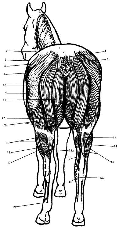
1.15 Muscles of the Horse, Rear View
(1) point of croup
(2) dock of tail
(3) point of hip or haunch (tuber coxae)
(4) levator muscles of tail
(5) depressor muscles of tail
(6) superficial gluteal muscles
(7) gluteal fascia
(8) tensor muscle of lateral femoral fascia
(9) biceps femoris muscle
(10) semitendinosus muscle
(11) semimembranosus muscle
(12) gracilis muscle
(13, 13a) gastrocnemius muscle and tendon
(14) soleus muscle
(15) lateral digital extensor muscle
(16, 16a) superficial digital flexor muscle and tendon
(17) deep digital flexor muscle
(18) inner or medial tendon of the cranial tibial muscle
(19) suspensory ligament
36
Equine Massage
Anatomy and Physiology of the Horse
37
t
le
ior par
h,
usc
vicis)
le
le
anc
usc
(continued)
le
usc
ior
ed from
usculus
usc
le
le
iv
rate m
le (poster
usculus ilio-
le
usculus spinalis
les
les
usc
usc
usc
le (m
usc
acis and cer
le (m
usc
lique m
lique m
extensor br
al ser
usc
le (m
usc
le