Breast Imaging: A Core Review (20 page)
Read Breast Imaging: A Core Review Online
Authors: Biren A. Shah,Sabala Mandava
Tags: #Medical, #Radiology; Radiotherapy & Nuclear Medicine, #Radiology & Nuclear Medicine

A. Category 0
B. Category 2
C. Category 3
D. Category 5
36a
A 50-year-old female presents for a screening mammogram.
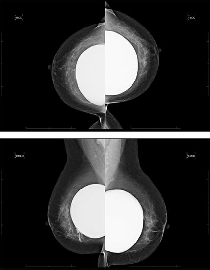
What is the salient finding?
A. Intracapsular rupture
B. Capsular calcification
C. Distortion of the implant contour
D. Free silicone with intracapsular and extracapsular rupture of the implant
36b
What is the BI-RADS assessment?
A. 0
B. 2
C. 3
D. 4
37
Which of the following is a high-risk lesion?
A. Peripheral duct papilloma
B. Intraductal papilloma
C. Intracystic papilloma
D. Papillary carcinoma in situ
38a
A 20-year-old female presenting with a new palpable abnormality in her right breast. Sonographic evaluation of the palpable area was performed.
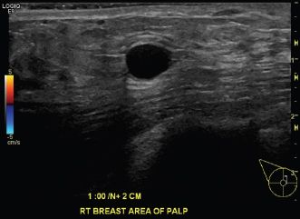
Which of the following is the best description of the sonographic finding?
A. Hypoechoic mass, smooth thin wall, sharp posterior border, posterior acoustic enhancement
B. Anechoic mass, smooth thin wall, sharp posterior border, posterior acoustic enhancement
C. Hypoechoic mass, smooth thin wall, sharp posterior border, increased through transmission
D. Anechoic mass, smooth thin wall, sharp posterior border, posterior acoustic shadowing
38b
What is the most likely diagnosis?
A. Solid mass
B. Complicated cyst
C. Complex cyst
D. Simple cyst
39
What is the most likely diagnosis for an encapsulated mass with “breast-within-a-breast” appearance on mammogram?
A. Fat necrosis
B. Fibroadenoma
C. Fibroadenolipoma
D. Galactocele
E. Lipoma
40
A 57-year-old female presents with a new palpable left breast mass that she states has grown rapidly over a period of <4 months. Her last screening mammogram was 6 months ago and interpreted as negative. Images from her most recent left breast diagnostic mammogram and ultrasound are shown.
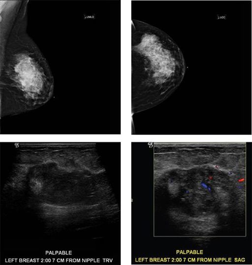
The most likely diagnosis is
A. Fibroadenoma
B. Hamartoma
C. Metaplastic carcinoma
D. Tubular adenoma
41
A 45-year-old female with a history type I diabetes presents with multiple palpable right breast masses that are firm on exam. Diagnostic imaging of the right breast was performed. Mammogram and ultrasound are shown.
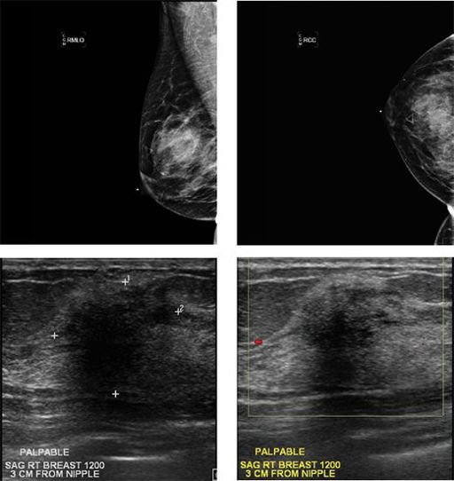
All of the masses were similar in appearance sonographically so a single mass was selected to sample under ultrasound guidance. Pathology demonstrates fibrous stromal proliferation and perivascular lymphocytic infiltrate consistent with diabetic mastopathy. The correct radiologic/pathologic correlation is
A. Concordant; excision recommended
B. Concordant; clinical follow-up recommended
C. Discordant; excision recommended
D. Discordant; repeat biopsy recommended
42
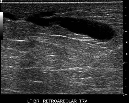
Shown below is a targeted ultrasound image of a 48-year-old female with clinical presentation of spontaneous left nipple yellow colored discharge. What is the most likely diagnosis?
A. Duct ectasia
B. Ductal carcinoma in situ
C. Papilloma
D. Papillary carcinoma
E. Paget disease of the nipple
43
Screening and diagnostic mammograms of a 52-year-old female demonstrate a spiculated mass with a central lucent area. Core biopsy of the mass proved it to be a radial scar. Subsequently, surgical excision was performed. What specific type of breast cancer may coexist with radial scar?
A. Ductal carcinoma in situ
B. Infiltrating lobular carcinoma
C. Inflammatory carcinoma
D. Medullary carcinoma
E. Tubular carcinoma
44
Approximately what percentage of all invasive breast malignancies are invasive lobular cancer?
A. <1%
B. 10%
C. 50%
D. 90%
45
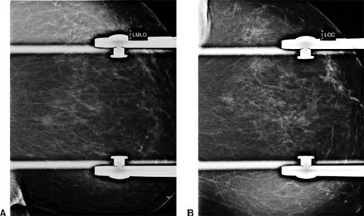
A 44-year-old female presents for additional views of an abnormality seen on screening mammography. Spot compression CC and MLO views are shown in images A and B. Prior comparison mammogram was negative in this location. Focused ultrasound was performed and demonstrates no sonographic abnormality. What is the most appropriate BI-RADS category given that the finding is not seen on ultrasound?
A. 0, incomplete, breast MRI recommended
B. 2, benign, return to screening in 1 year
C. 3, probably benign, 6-month follow-up mammogram recommended
D. 4, suspicious, biopsy is recommended
46
Which postconservation therapy change on MRI is considered a BI-RADS 4 finding and warrants tissue sampling to exclude recurrence?
A. Architectural distortion
B. Edema
C. Mass-like enhancement
D. Signal void or signal flare
E. Skin thickening
47
A 29-year-old female presents with a palpable mass in the right breast. The patient has ultrasound of the palpable lump. Findings are considered suspicious for malignancy. What is the recommended next step in evaluation of the suspicious mass?
A. MRI breast without and with contrast
B. Fine needle aspiration
C. Core biopsy
D. Diagnostic mammography
Other books
Messy Beautiful Love by Darlene Schacht
Uncle John’s Bathroom Reader Presents Flush Fiction by Bathroom Readers’ Institute
The Resurrection of Josephine by Melinda Barron
Condemned to Slavery by Bruce McLachlan
The Distinguished Guest by Sue Miller
Open Grave: A Mystery by Kjell Eriksson
The Pandora Directive: A Tex Murphy Novel by Aaron Conners
Hark! by Ed McBain
JaguarintheSun by Anya Richards
Heart Of The Wolf by Dianna Hardy