Oxford Handbook of Midwifery (66 page)
Read Oxford Handbook of Midwifery Online
Authors: Janet Medforth,Sue Battersby,Maggie Evans,Beverley Marsh,Angela Walker

- Emotional support
- Give verbal and non-verbal encouragement.
- Appreciate the woman’s individuality and needs.
- Be informative and gain consent for procedures.
 • Be aware of birth plan choices.
 - Communicate and give reassurance.
- Understand, appreciate, and be tolerant of her vocalizations, behaviour, and expressions during the second stage.
- Prepare her to welcome the baby into the world.
PRINCIPLES OF CARE IN THE SECOND STAGE OF LABOUR
275
Directed sustained versus spontaneous physiological pushing
Breath holding, closed glottis, and fixed diaphragm—commonly known as the Valsalva’s manoeuvre—is no longer recommended due to its negative effects (b see Breathing awareness, p. 246)
1,2
. At the peak of a contrac- tion the utero-placental blood flow ceases—a physiological factor that the fetus can cope with for short periods.This technique is said to reduce the length of the second stage, however, a technique for pushing infers that something needs to be done in place of the body’s own efforts, which undermines the mother’s ability and instincts.
1,2,3Spontaneous physiological pushing
This allows the woman’s body to tell her what to do. If she is left to push spontaneously, she will do so three to five times within a contraction, to coincide with extra surges. The length of pushing may be significantly reduced, but it is more intense and productive if the woman tunes into this natural pattern.
4Disadvantages
- Give verbal and non-verbal encouragement.
- Emotional support
- It is challenging for the woman to work with her own body and its intense sensations.
Advantages
- All fetal and maternal complications are minimized.
- Spiritual and psychological fulfilment/empowerment.
- Breathing awareness will automatically become easier and therefore assist with coping abilities.
- The woman can save her strength for the active/perineal phase.

- Bosomworth A, Bettany-Saltikov J (2006). Just take a deep breath… A review to compare the effects of spontaneous versus directed Valsalva pushing in the second stage of labour on maternal and fetal welbeing.
MIDIRS Midwifery Digest
16
(2), 157–65. - Byrom A, Downe S (2005). Second stage of labour: challenging the use of directed pushing.
Midwives
8
(4), 168–9. - Davies L (2006). Maternal pushing: to coach or not to coach.
Practising Midwife
9
(5), 38–40. - Perez-Botella M, Downe S (2006). Stories as evidence: why do midwives still use directed pushing.
British Journal of Midwifery
14
(10), 596–9.CHAPTER 14
Normal labour: second stage276
Care of the perineum
Care of the perineum is an important part of the birthing process, both for the midwife and the mother. Perineal trauma can have far-reaching effects, influencing the woman’s physical, emotional, and sexual relation- ships for the rest of her life. Perineal trauma is associated with:
- Urinary incontinence
- Faecal/flatus incontinence
- Dyspareunia
- Psychosocial factors as a result of the above
- A negative experience of motherhood.
Much of the routine care of the perineum is based on custom and practice; however, recent studies have shown how changes in management can help to enhance perineal integrity and minimize trauma.
Management
- Keeping the perineal area clean and free from faecal contamination is universally recognized.
- Conduct of the second stage of labour and perineal management should be discussed with the woman, to ensure informed choice and consideration of her birth plan.
- There is no evidence to suggest that applying hot flannels and pads to the perineum is beneficial; however, it may provide some temporary comfort for the woman if there are no objections to this practice.
- Antenatal massage of the perineum appears to be advantageous, however, there is no evidence to support massage of the perineum in the second stage of labour.
- Upright positions for birth generally enhance perineal integrity.
- There is no evidence to suggest that guarding the perineum is beneficial as opposed to hands-free management. There is a correlation between hands-free management and fewer episiotomies and third-degree tears.
- Women’s participation and confidence to breathe or pant the baby’s head out slowly, to gradually stretch the perineum, is beneficial.
- The midwife’s rapport and relationship with the woman and her birthing partner during labour is crucial, leading to less intervention in management of the perineum.
- Episiotomy should be reserved for fetal reasons only.
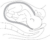
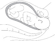
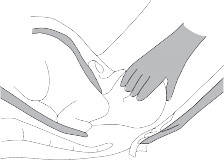
There has been much debate about whether applying pressure on the fetal head to maintain flexion, and the Ritgen manoeuvre to assist extension, are of value. Review of the key physiological principles would suggest that intervention using these techniques interferes with normal birthing mechanisms, by increasing the diameter of the fetal skull, thus
 inevitably leading to potential perineal trauma rather than preventing it.
This is best highlighted diagrammatically: see Fig. 14.2.
CARE OF THE PERINEUM
277
Flexion technique

Fig. 14.2a
The smallest diameter of the fetal head is maintained by an attitude of flexion. Further flexion applied by the birth attendant changes the position of the head to a larger diameter. This puts unnecessary pressure on the perineum, instead of its important role as a pivotal mechanism. Flexibility of the neck ensures the smallest diameter is maintained throughout the whole of the birth process.

Fig. 14.2b
The presenting part is halfway through the 90% curve of the birth canal. Partial extension occurs to maintain the smallest diameter (suboccipito-bregmatic). Extension of the head already occurs before it is visible therefore flexion at this point only maintains a larger diameter. As the birth canal is not straight there is no rationale for this technique.CHAPTER 14
Normal labour: second stage278
Ritgen manoeuvre

Fig. 14.2c
The Ritgen manoeuvre. As the fetal head emerges from the introitus, if over-extension is applied this results in the occipito-frontal diameter presenting – a considerably larger diameter. In an unassisted mechanism the head increasingly extends through the 90% angle so that it is able to utilize the resistance and pivotal force of the perineum and the pubic bone. This maintains the smallest diameter at the vaginal orifice.
This page intentionally left blank
CHAPTER 14
Normal labour: second stage280
Performing an episiotomy
In 1996 the World Health Organization (WHO) recommended an episi- otomy rate of less than 10% for normal deliveries. The only real indication for an episiotomy should be if the fetus is compromised in the second stage of labour and birth needs to be facilitated quickly. In rare cases a rigid perineum that is unquestionably obstructing the process of delivery may require episiotomy. Evidence suggests that:
1 - Protection against sphincter damage is unfounded
- Episiotomy may predispose to third-degree tears
- There is no difference between tears and episiotomies as far as pain and dyspareunia are concerned
- Episiotomy is more likely to cause unnecessary perineal trauma
- There is a risk of healing complications
- Episiotomy is contraindicated for third-degree tears, except in the event of shoulder dystocia, to prevent extensive perineal damage when undertaking internal manoeuvres to release the impacted fetal shoulder.
Awareness of anatomical structures is important, especially the location of the external sphincter muscle, which extends 2.5cm around the anus (see Fig. 14.3). A medio-lateral incision is used, as opposed to the midline inci- sion, which is associated with increased incidence of third-degree tears.
Performing an episiotomy
- Obtain consent after explaining the rationale determining the procedure.
- Prepare the necessary equipment:
- Sterile swabs
- Lidocaine 0.5% (10mL) or 1% (5mL)
- 10mL syringe
- Gauge 21 needle.
- Sterile swabs
- Clean the skin with antiseptic using aseptic technique.
- Insert the first and second fingers of your gloved hand into the vagina to protect the fetal skull from injury and anaesthetic.
- Episiotomy is usually performed on the right side. Insert the needle via the fourchette and direct it at a 45% angle from the midline for 4–5cm, to ensure a medio-lateral incision (Fig. 14.3).
- Withdraw the piston before infiltrating anaesthetic, to check that the needle is not in a blood vessel. If so, the needle must be withdrawn and repositioned.
- Urinary incontinence
- Bosomworth A, Bettany-Saltikov J (2006). Just take a deep breath… A review to compare the effects of spontaneous versus directed Valsalva pushing in the second stage of labour on maternal and fetal welbeing.
Other books
Vampire Coven Book 3: A Vampire's Embrace by C.L. Scholey
Bad Sisters by Rebecca Chance
The Night that Changed Everything by Anne McAllister
Training the Dom by d'Abo, Christine
Maxwell’s Reunion by M. J. Trow
Isle of Winds (The Changeling Series Book 1) by Fahy, James
Taco Noir by Steven Gomez
Sharing Space (The Complete Series) by Perez, Nina
Theatre by W Somerset Maugham
Kill the Dead by Richard Kadrey