Ross & Wilson Anatomy and Physiology in Health and Illness (110 page)
Read Ross & Wilson Anatomy and Physiology in Health and Illness Online
Authors: Anne Waugh,Allison Grant
Tags: #Medical, #Nursing, #General, #Anatomy

Figure 10.7
The cricoid cartilage.
The arytenoid cartilages
These are two roughly pyramid-shaped hyaline cartilages situated on top of the broad part of the cricoid cartilage forming part of the posterior wall of the larynx (
Fig. 10.8
). They give attachment to the vocal cords and to muscles and are lined with ciliated columnar epithelium.
Figure 10.8
A.
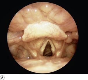
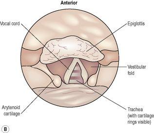
Bronchoscopic image of the open (abducted) vocal cords.
B.
Diagram of the vocal cords showing the principal structures.
The epiglottis
This is a leaf-shaped fibroelastic cartilage attached on a flexible stalk of cartilage to the inner surface of the anterior wall of the thyroid cartilage immediately below the thyroid notch (
Figs 10.4
and
10.8
). It rises obliquely upwards behind the tongue and the body of the hyoid bone. It is covered with stratified squamous epithelium. If the larynx is likened to a box then the epiglottis acts as the lid; it closes off the larynx during swallowing, protecting the lungs from accidental inhalation of foreign objects.
Ligaments and membranes
There are several ligaments that attach the cartilages to each other and to the hyoid bone (
Figs 10.5
,
10.6
and
10.8
).
Blood and nerve supply
Blood is supplied to the larynx by the superior and inferior laryngeal arteries and drained by the thyroid veins, which join the internal jugular vein.
The parasympathetic nerve supply is from the superior laryngeal and recurrent laryngeal nerves, which are branches of the vagus nerves. The sympathetic nerves are from the superior cervical ganglia, one on each side. These provide the motor nerve supply to the muscles of the larynx and sensory fibres to the lining membrane.
Interior of the larynx
The
vocal cords
are two pale folds of mucous membrane with cord-like free edges, which extend from the inner wall of the thyroid prominence anteriorly to the arytenoid cartilages posteriorly (
Fig. 10.8
).
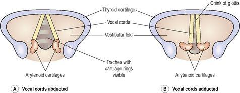
When the muscles controlling the vocal cords are relaxed, the vocal cords open and the passageway for air coming up through the larynx is clear; the vocal cords are said to be
abducted
(open,
Fig. 10.9A
). The pitch of the sound produced by vibrating the vocal cords in this position is low. When the muscles controlling the vocal cords contract, the vocal cords are stretched out tightly across the larynx (
Fig. 10.9B
), and are said to be
adducted
(closed). When the vocal cords are stretched to this extent, and are vibrated by air passing through from the lungs, the sound produced is high pitched. The pitch of the voice is therefore determined by the tension applied to the vocal cords by the appropriate sets of muscles. When not in use, the vocal cords are adducted. The space between the vocal cords is called the
glottis
.
Figure 10.9
The extreme positions of the vocal cords. A.
Abducted (open).
B.
Adducted (closed).
Functions
Production of sound
Sound has the properties of
pitch
,
volume
and
resonance
.
•
Pitch of the voice depends upon the
length
and
tightness
of the cords. Shorter cords produce higher pitched sounds. At puberty, the male vocal cords begin to grow longer, hence the lower pitch of the adult male voice.
•
Volume of the voice depends upon the
force
with which the cords vibrate. The greater the force of expired air, the more strongly the cords vibrate and the louder the sound emitted.
•
Resonance, or tone, is dependent upon the shape of the mouth, the position of the tongue and the lips, the facial muscles and the air in the paranasal sinuses.
Speech
This is produced when the sounds produced by the vocal cords are manipulated by the tongue, cheeks and lips.
Protection of the lower respiratory tract
During swallowing the larynx moves upwards, blocking the opening into it from the pharynx. In addition, the hinged epiglottis closes over the larynx. This ensures that food passes into the oesophagus and not into the trachea.
Passageway for air
The larynx links the pharynx above with the trachea below.
Humidifying, filtering and warming
These processes continue as inspired air travels through the larynx.
Trachea
Learning outcomes
After studying this section, you should be able to:
describe the location of the trachea
outline the structure of the trachea
explain the functions of the trachea in respiration.
Position
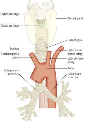
The trachea or windpipe is a continuation of the larynx and extends downwards to about the level of the 5th thoracic vertebra where it divides at the
carina
into the right and left primary bronchi, one bronchus going to each lung. It is approximately 10 to 11 cm long and lies mainly in the median plane in front of the oesophagus (
Fig. 10.10
).


