Ross & Wilson Anatomy and Physiology in Health and Illness (180 page)
Read Ross & Wilson Anatomy and Physiology in Health and Illness Online
Authors: Anne Waugh,Allison Grant
Tags: #Medical, #Nursing, #General, #Anatomy

Continued mobility of bone ends
Continuous movement results in fibrosis of the granulation tissue followed by fibrous union of the fracture.
Miscellaneous
These include:
•
infection (see below)
•
systemic illness
•
malnutrition
•
drugs, e.g. corticosteroids
•
ageing.
Complications of fractures
Infection (osteomyelitis,
p. 421
)
Pathogens enter through broken skin, although they may occasionally be blood-borne. Healing will not occur until the infection resolves.
Fat embolism
Emboli consisting of fat from the marrow in the medullary canal may enter the circulation through torn veins. They are most likely to lodge in the lungs and block blood flow through the pulmonary capillaries.
Axial skeleton
Learning outcomes
After studying this section you should be able to:
identify the bones of the skull (face and cranium)
list the functions of the sinuses and fontanelles of the skull
outline the characteristics of a typical vertebra
describe the structure of the vertebral column
explain the movements and functions of the vertebral column
identify the bones forming the thoracic cage.
The bones of the skeleton are divided into two groups: the
axial skeleton
and the
appendicular skeleton
(
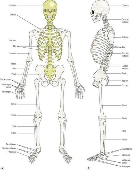
Fig. 16.9
).
Figure 16.9
The skeleton
. Axial skeleton in gold, appendicular skeleton in brown.
A.
Anterior view.
B.
Lateral view.
The axial skeleton consists of the
skull
,
vertebral column
,
ribs
and
sternum
. Together the bones forming these structures constitute the central bony core of the body, the axis.
Skull (
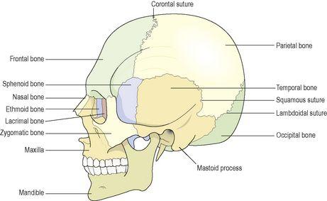
Figs 16.10
and
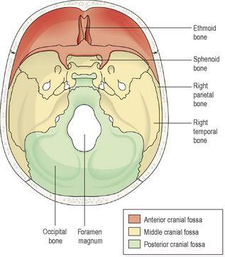
16.11
)
The skull rests on the upper end of the vertebral column and its bony structure is divided into two parts: the cranium and the face.
Figure 16.10
The bones of the skull and their sutures (joints).
Figure 16.11
The bones forming the base of the skull and the cranial fossae
– viewed from above.
Cranium
The cranium is formed by a number of flat and irregular bones that provide a bony protection for the brain. It has a
base
upon which the brain rests and a
vault
that surrounds and covers it. The periosteum lining the inner surface of the skull bones forms the outer layer of dura mater. In the mature skull the joints (
sutures
) between the bones are immovable (fibrous). The bones have numerous perforations (e.g. foramina, fissures) through which nerves, blood and lymph vessels pass. The bones of the cranium are:


