Ross & Wilson Anatomy and Physiology in Health and Illness (24 page)
Read Ross & Wilson Anatomy and Physiology in Health and Illness Online
Authors: Anne Waugh,Allison Grant
Tags: #Medical, #Nursing, #General, #Anatomy

Functions of the thoracic cage
The functions of the thoracic cage are:
•
It protects the thoracic organs including the heart, lungs and large blood vessels.
•
It forms joints between the upper limbs and the axial skeleton. The upper part of the sternum, the
manubrium
, articulates with the clavicles forming the only joints between the upper limbs and the axial skeleton.
•
It gives attachment to the muscles of respiration:
–
intercostal muscles
occupy the spaces between the ribs and when they contract the ribs move upwards and outwards, increasing the capacity of the thoracic cage, and inspiration occurs
–
the
diaphragm
is a dome-shaped muscle which separates the thoracic and abdominal cavities. It is attached to the bones of the thorax and when it contracts it assists with inspiration.
•
It enables breathing to take place.
Appendicular skeleton
The appendicular skeleton consists of the shoulder girdles and upper limbs, and the pelvic girdle and lower limbs (
Fig. 3.27
).
The shoulder girdles and upper limbs
Each shoulder girdle consists of a clavicle and a scapula. Each upper limb comprises:
•
1 humerus
•
1 radius
•
1 ulna
•
8 carpal bones
•
5 metacarpal bones
•
14 phalanges.
The pelvic girdle and lower limbs
The bones of the pelvic girdle are the two innominate bones and the sacrum. Each lower limb consists of:
•
1 femur
•
1 tibia
•
1 fibula
•
1 patella
•
7 tarsal bones
•
5 metatarsal bones
•
14 phalanges.
Functions of the appendicular skeleton
The appendicular skeleton has two main functions.
•
Voluntary movement
. The bones, muscles and joints of the limbs are involved in movement of the skeleton. This ranges from very fine finger movements needed for writing to the coordinated movement of all the limbs associated with running and jumping.
•
Protection of delicate structures
. Blood vessels and nerves lie along the length of bones of the limbs and are protected from injury by the associated muscles and skin. These structures are most vulnerable where they cross joints and where bones can be felt immediately below the skin.
Cavities of the body
The organs that make up the systems of the body are contained in four cavities: cranial, thoracic, abdominal and pelvic.
Cranial cavity
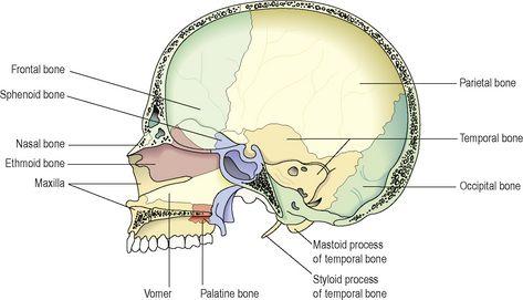
The cranial cavity contains the brain, and its boundaries are formed by the bones of the skull (
Fig. 3.31
):
Anteriorly
– 1 frontal bone
Laterally
– 2 temporal bones
Posteriorly
– 1 occipital bone
Superiorly
– 2 parietal bones
Inferiorly
– 1 sphenoid and 1 ethmoid bone and parts of the frontal, temporal and occipital bones.
Figure 3.31
Bones forming the right half of the cranium and the face – viewed from the left.
Thoracic cavity
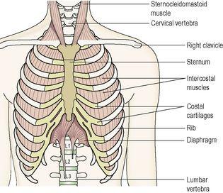
This cavity is situated in the upper part of the trunk. Its boundaries are formed by a bony framework and supporting muscles (
Fig. 3.32
):
Anteriorly
– the sternum and costal cartilages of the ribs
Laterally
– 12 pairs of ribs and the intercostal muscles
Posteriorly
– the thoracic vertebrae
Superiorly
– the structures forming the root of the neck
Inferiorly
– the diaphragm, a dome-shaped muscle.
Figure 3.32
Structures forming the walls of the thoracic cavity and associated structures.
Contents
The main organs and structures contained in the thoracic cavity are shown in
Figure 5.10, page 79
. These include:
•
the trachea, 2 bronchi, 2 lungs
•
the heart, aorta, superior and inferior vena cava, numerous other blood vessels
•
the oesophagus
•
lymph vessels and lymph nodes
•
some important nerves.
The
mediastinum
is the name given to the space between the lungs including the structures found there, such as the heart, oesophagus and blood vessels.
Abdominal cavity
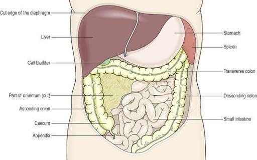
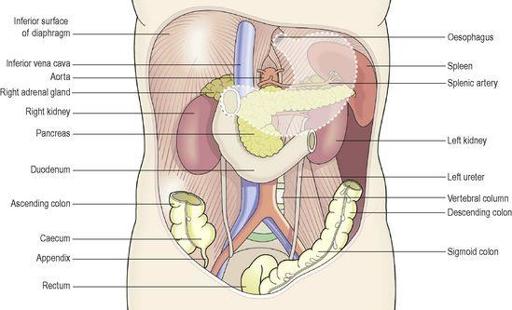
This is the largest cavity in the body and is oval in shape (
Figs 3.33
and
3.34
). It is situated in the main part of the trunk and its boundaries are:
Superiorly
– the diaphragm, which separates it from the thoracic cavity
Anteriorly
– the muscles forming the anterior abdominal wall
Posteriorly
– the lumbar vertebrae and muscles forming the posterior abdominal wall
Laterally
– the lower ribs and parts of the muscles of the abdominal wall
Inferiorly
– it is continuous with the pelvic cavity.
Figure 3.33
Organs occupying the anterior part of the abdominal cavity and the diaphragm (cut).
Figure 3.34
Organs occupying the posterior part of the abdominal and pelvic cavities.
The broken line shows the position of the stomach.
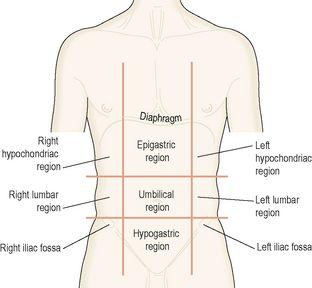
By convention, the abdominal cavity is divided into the nine regions shown in
Figure 3.35
. This facilitates the description of the positions of the organs and structures it contains.
Figure 3.35
Regions of the abdominal cavity.
Contents
Most of the abdominal cavity is occupied by the organs and glands of the digestive system (
Figs 3.33
and
3.34
). These are:
•
the stomach, small intestine and most of the large intestine
•
the liver, gall bladder, bile ducts and pancreas.
Other structures include:
•
the spleen
•
2 kidneys and the upper part of the ureters
•
2 adrenal (suprarenal) glands
•
numerous blood vessels, lymph vessels, nerves
•
lymph nodes.
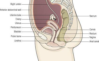
Pelvic cavity
The pelvic cavity is roughly funnel shaped and extends from the lower end of the abdominal cavity (
Figs 3.36
and
3.37
). The boundaries are:
Superiorly
– it is continuous with the abdominal cavity
Anteriorly
– the pubic bones
Posteriorly
– the sacrum and coccyx
Laterally
– the innominate bones
Inferiorly
– the muscles of the pelvic floor.
