Brain on Fire (23 page)
Authors: Susannah Cahalan

This was not what my mother wanted to hear. Cancer had always been the greatest fear, the word she dared not utter. Now this doctor was tossing it off casually as part of a bet.

Meanwhile, two plastic tubes, placed securely in Styrofoam boxes, had arrived at the University of Pennsylvania, transported in a refrigerator in the back of a FedEx truck; one contained transparent cerebrospinal fluid, as clear as unfiltered water, and another held blood, which started to look like dehydrated urine as, over time, the red blood cells dropped to the bottom. The test tubes were coded 0933, labeled with my initials, SC, and placed in a negative-80 degree freezer waiting for the lab to conduct its tests. They were addressed to the lab run by neuro-oncologist Dr. Josep Dalmau, whom Dr. Najjar had mentioned during his first visit and whom Dr. Russo had since e-mailed to ask if he would take a look at my case.
Four years earlier, in 2005, Dr. Dalmau had been the senior author on a paper in the neuroscience journal
Annals of Neurology
that focused on four young women who had developed prominent psychiatric symptoms and encephalitis. All had white blood cells in their cerebrospinal fluid, confusion, memory problems, hallucinations, delusions, and difficulty breathing, and they all had tumors called teratomas in their ovaries. But the most remarkable finding was that all four patients had similar antibodies that appeared to be reacting against specific areas of the brain, mainly the hippocampus. Something about the combination of the tumor and the antibodies was making these women very sick.
Dr. Dalmau had noticed a pattern in these four women; now he had to learn more about the antibody itself. He and his research team began to work night and day on an elaborate immunohistrochemistry experiment involving frozen sections of rat brains, which had been sliced into paper-thin pieces and then exposed to the cerebrospinal fluid of those four sick women. The hope was that the antibodies from the cerebrospinal fluid would bind directly to some receptors in the rat brain and reveal a characteristic design. It took eight months of tinkering before a pattern finally emerged.
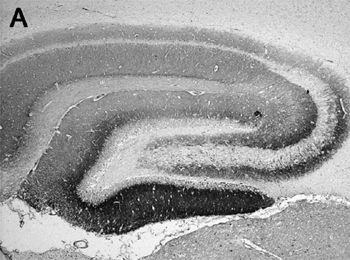
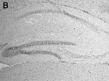
Dr. Dalmau had prepared the rat brain slides all the same, placing a small amount of cerebrospinal fluid from each of the four patients on each. Twenty-four hours later:

A: Section of rat brain in the hippocampus that shows the reactivity of the cerebrospinal fluid of a patient with anti-NMDAR encephalitis. The brown staining corresponds to the patient’s antibodies that have bound to the NMDA receptors.

B: A similar section of hippocampus of an individual without NMDA receptor antibodies.
Four beautiful images, like cave drawings or abstract seashell patterns, revealed the antibodies’ binding to the naked eye. “It was a moment of great excitement,” Dr. Dalmau later recalled. “Everything had been negative. Now we became totally positive that all four not only had the same illness, but the same antibody.”
He had clarified that the pattern of reactivity was more intense in the hippocampus of the rat brain, but this was only the beginning. A far more difficult question now arose: Which receptors were these antibodies targeting? Through a combination of trial and error, plus a few educated guesses about which receptors are most common in the hippocampus, Dr. Dalmau and his colleagues eventually identified the target. Using a kidney cell line bought from a commercial lab that came with no receptors on their surfaces at all, a kind of “blank slate,” his lab introduced DNA sequences that direct the cells to make certain types of receptors, allowing the lab to control which receptors were available for binding. Dalmau chose to have them express only NMDA receptors, after figuring out that those were the most likely to have been present in high volume in the hippocampus. Sure enough, the antibodies in the cerebrospinal fluid of the four patients bound to the cells. There was his answer: the culprits were NMDA-receptor-seeking antibodies.
NMDA (N-methyl-D-aspartate acid) receptors are vital to learning, memory, and behavior, and they are a main staple of our brain chemistry.
39
If these are incapacitated, mind and body fail. NMDA receptors are located all over the brain, but the majority are concentrated on neurons in the hippocampus, the brain’s primary learning and memory center, and in the frontal lobes, the seat of higher functions and personality. These receptors receive instructions from chemicals called neurotransmitters. All neurotransmitters carry only one of two messages: they can either “excite” a cell, encouraging it to fire an electrical impulse, or “inhibit” a cell, which hinders it from firing. These simple conversations between neurons are at the root of everything we do, from sipping a glass of wine to writing a newspaper lead.
In those unfortunate patients with Dr. Dalmau’s anti-NMDA-receptor encephalitis, the antibodies, normally a force for good in the body, had become treasonous persona non grata in the brain. These receptor-seeking antibodies planted their death kiss on the surface of a neuron, handicapping the neuron’s receptors,
making them unable to send and receive those important chemical signals. Though researchers are far from fully understanding how NMDA receptors (and their corresponding neurons) affect and alter behavior, it’s clear that when they are compromised the outcome can be disastrous, even deadly.
Still, a few experiments have offered up some clues as to their importance. Decrease NMDA receptors by, say, 40 percent, and you might get psychosis; decrease them by 70 percent, and you have catatonia. In “knockout mice” without NMDA receptors at all, even the most basic life functions are impossible: most die within ten hours of birth due to respiratory failure.
40
Mice with a very small number of NMDA receptors don’t learn to suckle, and they simply starve to death within a day or so. Those mice with at least 5 percent of their NMDA receptors intact survive, but exhibit abnormal behavior and strange social and sexual interactions. Mice with half their receptors in working order also live, but they show memory deficits and abnormal social relationships.
As a result of this additional research, in 2007, Dr. Dalmau and his colleagues presented another paper, now introducing his new class of NMDA-receptor-seeking diseases to the world. This second article identified twelve women with the same profile of neurological symptoms, which could now be called a syndrome.
41
They all had teratomas, and almost all of them were young women. Within a year after publication, one hundred more patients had been diagnosed; not all of them had ovarian teratomas and not all of them were young women (some were men and many were children), enabling Dr. Dalmau to do an even more thorough study on the newly discovered, but nameless, disease.
“Why not name it the Dalmau disease?” people often asked him. But he didn’t think “Dalmau disease” sounded right, and it was no longer customary to name a disease after its discoverer. “I didn’t think that would be wise. It’s not very humble.” He shrugged.
By the time I was a patient at NYU, Dr. Dalmau had fine-tuned his approach, designing two tests that could swiftly and accurately diagnose the disease. As soon as he received my samples, he could
test the spinal fluid. If he found that I had anti-NMDA-receptor autoimmune encephalitis, it would make me the 217th person worldwide to be diagnosed since 2007. It just begged the question: If it took so long for one of the best hospitals in the world to get to this step, how many other people were going untreated, diagnosed with a mental illness or condemned to a life in a nursing home or a psychiatric ward?
RHUBARB
B
y my twenty-fifth day in the hospital, two days after the biopsy, with a preliminary diagnosis in sight, my doctors thought it was a good time to officially assess my cognitive skills to record a baseline. This test would be a fulcrum, a turning point, that would measure what kind of progress they could expect in the future through the various stages of treatment. Beginning on the afternoon of April 15, a speech pathologist and a neuropsychologist visited me for two days in a row, each for separate assessments.
The speech pathologist, Karen Gendal, did the first assessment, starting with basic questions: “What’s your name?” “How old are you?” “Are you a woman?” “Do you live in California?” “Do you live in New York?” “Do you peel a banana before eating it?” and so forth. I was able to answer all of these questions, though I did so slowly. But when she asked the more open-ended question “Why are you in the hospital?” I could not explain. (To be fair, the doctors didn’t know either, but I could not provide even the basics.)
After some spotty and tangential answers, I finally said, “I can’t get my ideas from my head out.” She nodded: this was a typical response for people suffering from aphasia, a language impairment related to brain injury. I also had something called dysarthria, a motor speech impairment caused by a weakness in the muscles of the face, throat, or vocal cords.
Gendal asked me to stick out my tongue, which trembled from the effort. It had a reduced range of motion on both sides, which was contributing to my inability to articulate.
“Would you smile for me?”
I tried, but my facial muscles were so weak that no smile came. She wrote down “hypo-aroused,” a medical term for lethargic, and also noted that I was not fully alert. When I did talk, the words came out without any emotional register.
She moved on to cognitive abilities. Holding up her pen, she asked, “What is this?”
“Ken,” I answered. This, again, wasn’t too unusual for someone at my level of impairment. They call it
phonemic paraphasia,
where you substitute one word for another that sounds similar.
When she asked me to write my name, I painstakingly drew out an “S,” tracing the letter multiple times before moving on to the “U,” where I did the same. It took several minutes for me to write my name. “Okay, would you write this sentence out for me: ‘Today is a nice day.’”
I drew out the letters, retracing each of them several times and misspelling some words. My handwriting was so poor that Gendal could hardly make out the sentence.
She wrote her impression in the chart: “It is difficult to determine at just two days post-operation to what extent communication deficits are language-based versus medication or cognitive. Clearly communication function is dramatically reduced from pre-morbid level when this patient was working as a successful journalist for a local paper.” In other words, there seemed to be a dramatic change between the person I had been and the person I was now, but it was difficult at the time to distinguish my problems understanding from my inability to communicate—and whether these would persist in the long or short term.