Equine Massage: A Practical Guide (7 page)
Read Equine Massage: A Practical Guide Online
Authors: Jean-Pierre Hourdebaigt

18
Equine Massage
vagina, and external genitalia. Proper fluid circulation and relaxation of the nervous system will ensure peak performance for reproduction purposes.
The Skeletal System
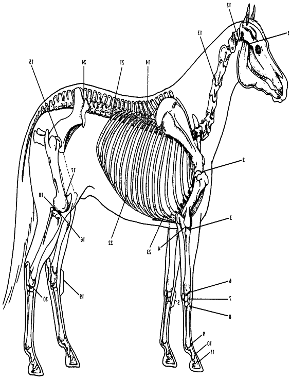
The skeletal system serves as a framework for the horse’s body, giving the muscles something to work against, and defining the animal’s overall size and shape. The skeleton also protects the horse’s vital internal tissues and organs. For example, the skull protects the brain; the rib cage protects the lungs and heart; the vertebral column protects the spinal cord.The skeleton is made up of over 200
bones.
Bones
Bones vary in size and shape according to their function.With the exception of the enamel-covered teeth, bones are the body’s hard-est substances and can withstand great compression, torque, and tension. A tough membrane called the
periosteum
covers and protects the bones and provides for the attachment of the joint capsules, ligaments, and tendons. Injury to the periosteum may result in undesirable bone growths such as splints, spavin, and ringbone.
Bones are held together by
ligaments
; muscles are attached to the bones by
tendons
.
The articulating surface of the bone is covered with a thick, smooth cartilage that diminishes concussion and friction.
Long bones are found in the limbs; short bones in the joints; flat bones in the rib cage, skull, and shoulder; and irregularly shaped bones in the spinal column and limbs.
1.6 A Bone
(1) periosteum
(4) spongy bone with marrow cavities
(2) compact bone
(5) epiphyseal plate
(3) medullary cavity
(6) articulate hyaline cartilage
Anatomy and Physiology of the Horse
19
The
long bones
of the limbs (humerus, radius, femur, tibia, cannon bones) function mainly as levers and aid in the support of weight.
Short bones,
found in complex joints such as the knee (carpus), hock (tarsus), and ankle (fetlock), absorb concussion.
Flat bones
protect and enclose the cavities containing vital organs: skull (brain) and ribs (heart and lungs). Flat bones also provide large areas for the attachment of muscles.
Components of the skeleton of the horse are as follows:
❖ The skull consists of 34 irregularly shaped bones.
❖ The spine consists of 7 cervical vertebrae, 17 to 19 thoracic vertebrae (usually 18), 5 to 6 lumbar vertebrae (sometimes fused together), and 5 fused sacral vertebrae (the sacrum).
The tail consists of 18 coccygeal vertebrae, although this number can vary considerably.
❖ The rib cage consists of 18 pairs (usually) of ribs springing from the thoracic vertebrae, curving forward and meeting at the breastbone (sternum).
❖ The forelegs carry 60 percent of the horse’s body weight.
Comprising the forelegs are the shoulder blade (scapula), humerus, radius, knee (8 carpal bones), cannon, splints, long and short pasterns, and the pedal (or coffin) bone.
❖ Comprising the hind legs are the pelvis (ilium, ischium, pubis), femur, tibia and fibula, the tarsus or hock (7 bones), cannons and splints, pasterns (long and short), and the pedal (or coffin) bone.
The Joints
Joints are the meeting places between two bones. Movement of the horse is dependent upon the contraction of muscles and the corresponding articulation of the joints.
Some joints in the horse’s body are not movable, but most are and permit a great range of motion.
The ends of the bones are lined with hyaline cartilage, which provides a smooth surface between the bones and acts as a shock absorber when compressed—for example, during takeoff and landing while jumping, and for torque during quick turns.
The
joint capsule,
also known as the capsular ligament, is sealed by the synovial membrane, which produces a viscous, lubricating secretion, the synovial fluid.

20
Equine Massage
Anatomy and Physiology of the Horse
21
al joint
al joint
al joint
tebr
nal joint
er
hondr
k joint (tarsus)
(17) femoropatellar component of stifle joint
(18) femorotibial component of stifle joint
(19) hoc
(20) talocrur
(21) costov
(22) costoc
(23) costoster
(24) sacroiliac joint
al disc joint
al disc joint
vertebr
vertebr
n joint
k joint
acic inter
vical inter
(9) fetloc
(10) paster
(11) coffin joint
(12) atlanto-occipital joint
(13) cer
(14) thor
(15) hip joint
(16) stifle joint
ular joint [TMJ])
ist)
pus or wr
pal joint
adial component of elbow joint
pal joint
pal joint
pometacar
Joints of the Horse
adiocar
1.7
(1) jaw joint (temporomadib
(2) shoulder joint
(3) humeror
(4) humeroulnar component of elbow joint
(5) knee joint (car
(6) r
(7) intercar
(8) car


22
Equine Massage
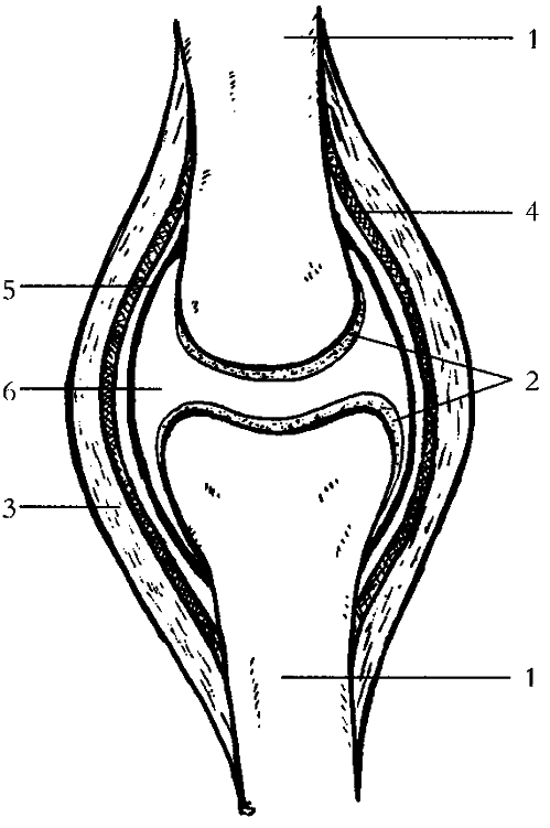
1.8 A Joint
(1) bone
(2) hyaline cartilage
(3) ligament
(4) fibrous capsule
(5) synovial lining
(6) joint cavity (with synovial fluid)
The Ligaments
A ligament is a band of connective tissue that links one bone to another (
tendons
connect muscles to bones). Ligaments are made up of collagen fiber, a fibrous protein found in the connective tissue. Ligaments have a limited blood supply. Consequently, if a ligament is injured, say by a sprain, it tends to heal slowly and sometimes incorrectly.
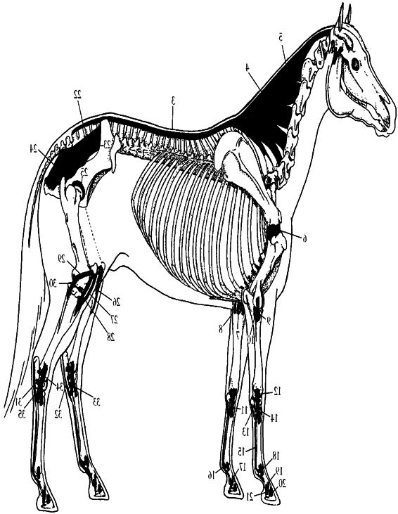
Most ligaments are located around joints to give extra support (capsular ligaments and collateral ligaments) or to prevent an excessive or abnormal range of motion and to resist the pressure of lateral torque (a twisting motion). (See figure 1.10, page 24.) The horse experiences lateral torque when turning sharply.
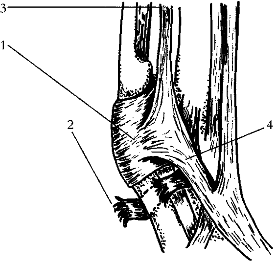
1.9 Ligaments of the Fetlock
Joint
(1) palmar annular ligament, which
provides strong support
(2) example of a torn ligament, the
digital annular ligament
(3) suspensory ligament (superior
sesamoidean part)
(4) suspensory ligament extension to
common digital extensor tendon
Anatomy and Physiology of the Horse
23
Ligaments have little contracting power and therefore must work in conjunction with muscle action.Within very narrow limits, ligaments are somewhat elastic but are inflexible enough to offer support in normal joint play. If overstretched or repeatedly stretched, a ligament might lose up to 25 percent of its strength.
Such a ligament may need surgical stitching to recover its full tensile strength. Severe ligament sprain will lead to joint instability.
Several ligamentous structures help support and protect the vertebral column, pelvis, neck, and limbs from suddenly imposed strain.
The Muscular System
There are three classes of muscles: smooth, cardiac, and skeletal.
The
smooth
and
cardiac muscles
are involuntary, or autonomic; they play a part in the digestive, respiratory, circulatory, and urogenital systems.
For the most part,
skeletal muscles
are voluntary; they function in the horse’s movements. In massage, we are concerned with the more than 700 skeletal muscles that are responsible for the movement of the horse.
There are two types of skeletal muscle fibers: slow twitch fibers (ST) and fast twitch fibers (FT).
Slow twitch fibers
are aerobic fibers; they need oxygen in order to do their job.Thus ST fibers require a good supply of blood to bring oxygen to them and to remove waste products created during exercise. ST fibers have strong endurance qualities.
Fast twitch fibers
are anaerobic fibers; they do not need oxygen to work and therefore are able to deliver the quick muscular effort required for a sudden burst of speed. However, FT fibers are only able to perform for short periods of time.
The ratio of ST to FT fibers is genetically inherited. Careful selective breeding can emphasize these features in a horse. For example, the muscles of the Quarter Horse are mostly FT fibers, and the breed is noted for its dazzling sprint. On the other hand, a heavy horse, such as a draft horse, has more ST fibers and is noted for its strength and endurance abilities. Whether we are talking about FT or ST fibers, a muscle is made up of a fleshy part and two tendon attachments.The fleshy part, or
muscle belly,
is the part that contracts in response to nervous command. During contraction, the muscle fibers basically fold on themselves, shortening the fibers and resulting in muscle movement. The muscle belly is made up of many muscle fibers arranged in bundles, with each bundle wrapped in connective tissue (
fascia
). The fascia covers, supports, and separates the muscle bundles and the whole muscle

24
Equine Massage
Anatomy and Physiology of the Horse
25
al ligament
al ligament of stifle joint
al ligament of tarsal joint
al ligament of tarsal joint
hes of medial collater
dle patellar ligament
al patellar ligament
al femoropatellar ligament