Equine Massage: A Practical Guide (8 page)
Read Equine Massage: A Practical Guide Online
Authors: Jean-Pierre Hourdebaigt

al collater
al collater
anc
ligament)
(24) sacrosciatic ligament
(25) capsular ligament
(26) medial patellar ligament
(27) mid
(28) later
(29) later
(30) later
(31) ligament connecting talus and calcaneus
(32) br
(33) medial collater
(34) later
(35) calcaneometa tarsal ligament (plantar
k joint
n joint
ior
ed fromiv
pal joints
le)
usc
ior sesamoidean ligaments
al ligament of fetloc
al sesamoidean ligament
al ligament of paster
al ligament of coffin joint
y ligament (super
y ligament of navicular bone
k joint)
al collater
al collater
al collater
al sacroiliac ligament
sesamoidean ligament der
interosseous m
(fetloc
(14) dorsal ligaments of car
(15) suspensor
(16) distal or infer
(17) medial collater
(18) later
(19) later
(20) later
(21) suspensor
(22) dorsal sacroiliac ligament
(23) later
pal joint
pal joint
pal bone
y car
hal ligament
hal ligament
adioulnar ligament
adioulnar ligament
al ligament of car
al ligament of car
ior) ligament of jaw joint
al ligament of elbow joint
erse r
t of nuc
erse r
al ligament of elbow joint
t of nuc
ansv
ansv
aments of the Horse
al tr
al collater
al ligament of jaw joint
aspinous ligament
al collater
Lig
1.10
(1) later
(2) caudal (poster
(3) supr
(4) funicular par
(5) lamellar par
(6) capsular ligament of shoulder joint
(7) medial collater
(8) medial tr
(9) later
(10) later
(11) medial collater
(12) later
(13) distal ligament of accessor
26
Equine Massage
itself. This arrangement allows for greater support, strength, and flexibility in the movement between each of the muscle groups.
Tendons
The tendon is the portion of the muscle that attaches to the bone.
It is made up of
connective tissue
—a dense, white, fibrous tissue much like that of a ligament. The
origin tendon
is the tendon that attaches the muscle to the least movable bone; the
insertion tendon
attaches the muscle to the movable bone, so that on contraction the insertion is brought closer to the origin.Tendons attach to the periosteum of the bone; the fibers of the tendon blend with the periosteum fibers because of their similar collagen make-up.
Tendons can be fairly short, or quite long as is seen with the flexor and extensor muscles of the lower legs. Usually, tendons are rounded but they can be flattened like the tendons that attach along the spine.
Because of their high-tensile strength, tendons can endure an enormous amount of tension, usually more than the muscle itself can produce; consequently, tendons do not rupture easily.They are not as elastic as muscle fibers, but they are more elastic than ligament fibers.
Tendons can “stress up” after heavy exercise, meaning that they can stay contracted. Gentle massage and stretching exercises will loosen residual tension. (See the neuromuscular technique in chapter 5.)Inflamed tendons are at great risk of being strained or overstretched.The horse has no muscles below the knee or hock; consequently, many leg muscles have long tendons that run down the legs over the joints.These tendons are protected by sheaths, or tendon bursae. Chronic irritation of the sheath can result in excess fluid production and soft swellings. Cold hydrotherapy (chapter 4) and massage will help increase circulation and keep inflammation down. If the inflammation persists, check with your veterinarian.
Muscles
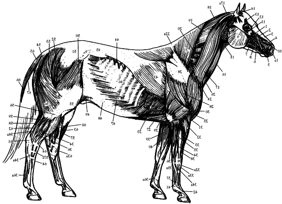
Muscles come in all shapes and sizes. Some are small, some are large; some are thin, some are bulky. Look at the muscle charts to note the variety of shapes in the horse’s muscle structure.
Muscles act together to give the horse his grace and power.
Muscles work in three different ways: isometric contraction, concentric contraction, and eccentric contraction.
Isometric contraction
occurs when a muscle contracts without causing any movement. During standing, for example, isometric contraction ensures stability.


Anatomy and Physiology of the Horse
27
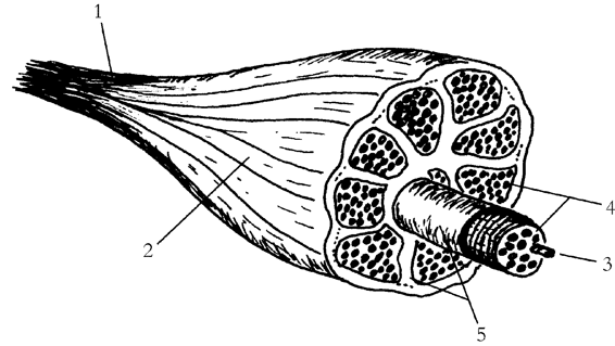
1.11 Cross-Section of a Skeletal Muscle
(1) tendon
(2) muscle belly
(3) muscle fiber (containing thick and thin filaments)
(4) bundles (made up of fibers)
(5) fascia
Concentric contraction
occurs when a muscle shortens as it contracts, causing articular movements. Concentric contraction is mostly seen in regular movements such as
protraction
(forward movement) or
retraction
(backward movement) of the limbs, and in any movement of the neck or back.
Eccentric contraction
occurs when a muscle gradually releases as it elongates. Eccentric contraction assists regular movements to avoid jerky, unstable actions; it also plays a role in shock absorption during the landing phase of jumping.
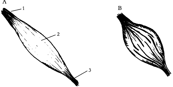
1.12 A Muscle
(A) Relaxed
(B) Contracted
[1] origin tendon
[2] muscle belly
[3] insertion tendon

28
Equine Massage
Anatomy and Physiology of the Horse
29
ing
ing
le
ascia)
le
le
le
TFL)
usc
usc
usc
le
usc
or ,
(continued)
usc
ae m
ior
y ligament,
t of hamstr
t of hamstr
rate m
acolumbar f
le
ascia of thigh
lique m
le
le
usc
le in fold of flank
al f
usc
le (par
le (par
iceps sur
usc
usc
asciae latae
usc
usc
ascia
h of suspensor
ascia (thor
ascia
le of later
is m
al f
y ligament (super
anc
t of dorsal ser
al f
us dorsi m
hment)
usc
ascia
hing to common digital extensor tendon
nal intercostal m
nal abdominal ob
al femor
al crur
al head of gastrocnemius m
usculus tensor f
hilles tendon of tr
sesamoidean section)
attac
of attac
(m
group)
group)
57a) long digital extensor m
Ac
(41) suspensor
(42) extensor br
(43) latissim
(44) caudal par
(45) lumbodorsal f
(46) exter
(47) exter
(48) aponeurosis (broad and flattened tendon
(49) remains of skin m
(50) tensor m
(51) gluteal f
(52) superficial gluteal m
(53) biceps femor
(54) semitendinosus m
(55) later
(56) later
(57,
(58)
(58a) later
t
t
ior
le
le and
ior par
ior par
usc
le and
usc
le and
le (anter
k
usc
usc
le and its
usc
usc
le and tendon
le
rate m
le (anter
t
le (poster
usc
usc
le
le
usc
al m
al)
usc
usc
usc
al ser
le of nec
al m
usc
al m
entr
le
iceps
le
pal flexor m
al)
al)
iceps
pal extensor m
long and shor
t of v
usc
usc
,
al digital extensor m
al car
pal extensor m
pal flexor m
pal flexor m
adial car
acic par
t of superficial pector
al head of tr
hialis m
dle car
anial deep pector
anial superficial pector
ac
lique car
of deep pector
par
of deep pector
32a) r
tendon
33a) common digital extensor m
tendon
34a) later
tendon
35a) later
two tendons
36a) deep digital flexor m
(23) thor
(24) cr
(25) cr
(26) remains of skin m
(27) caudal deep pector
(28) deltoideus m
(29) long head of tr
(30) later
(31) br
(32,
(33,