Life on a Young Planet (25 page)
Read Life on a Young Planet Online
Authors: Andrew H. Knoll

The eukaryote-rich rocks of the Doushantuo Formation, then, are much younger than the Great Wall dolomites, and they postdate the fossiliferous rocks of Spitsbergen, as well. Indeed, these rocks are only 50 to 60 million years older than the Cambrian cliffs along the Kotuikan River.
1
Perhaps, then, Doushantuo fossils will not only illuminate the rise of nucleated organisms but hint, as well, of further biological transformation about to begin.
How do we identify a fossil as eukaryotic? For plants and animals, the distinction is easy—no bacteria or archaeans build anything like a leaf or a shell. Microscopic fossils, however, can be more challenging. Biologists find it easy to distinguish between prokaryotes and eukaryotes based on myriad features of cellular organization, genetics, and physiology, but none of these features is available to paleontologists. We have to rely on form.
When Precambrian research was young, paleontologists tried hopefully to recognize eukaryotic microfossils on the basis of size or preserved features of cell biology. Neither worked. It is easy to see the attraction of size—on average, eukaryotic cells are larger than bacteria, and diameter is simple to measure. At the extremes of the scale, size can indeed be informative—bacterial cells more than a millimeter long are unknown, and neither do we know of eukaryotes only 300 nanometers across.
2
But at intermediate sizes (commonly encountered in Proterozoic rocks) there is strong overlap between bacteria and eukaryotes. Tiny green algae in the open ocean are less than a micron in diameter, and—
recalling those cigar-shaped fossils in Kotuikan cherts—the resting cells of cyanobacteria can be well over 100 microns long.
3
Cyanobacteria that form extracellular envelopes complicate the picture further, because a colony of 10-micron cells can be enclosed in a preservable coat 100 microns across.
If size isn’t foolproof, what about preserved details of cell biology? One of the first Proterozoic fossil assemblages to be discovered was that of the 830–810-million-year-old Bitter Springs Formation in central Australia. Bitter Springs fossils occur in chert nodules within carbonates deposited in ephemeral lakes on an arid coastal plain. The cherts contain beautiful cyanobacteria described by UCLA’s Bill Schopf, as well as simple spherical fossils about 10 microns in diameter. Some of these spheres are hollow and were originally interpreted as cyanobacteria. Others, although essentially identical, contain small, dark inclusions of organic matter and so were interpreted as eukaryotic algae with preserved nuclei. Nuclei are mostly water, along with highly nutritious proteins and nucleic acids. In consequence, they are quickly and completely obliterated soon after death—so completely that in the entire fossil record only a handful of plausible fossil nuclei have ever been identified. On the other hand, decomposing cyanobacteria and algae commonly contain small balls of organic matter formed as cell contents shrivel. Decaying cytoplasm provides a satisfactory explanation for the “black spots” in Bitter Springs and other Proterozoic microfossils. Some of these fossils may be eukaryotic, but (unlike leopards) we can’t tell them by their spots.
What really makes eukaryotic fossils stand out is
morphology
. In
chapter 3
, we noted that some cyanobacteria have cell shapes and colony forms not duplicated by other bacteria. Similarly, some (but not all) eukaryotic cells display features that are unknown in prokaryotic organisms. Doushantuo fossils illustrate how paleontologists recognize and interpret early fossil eukaryotes.
In and around the Yangtze Gorges, chert nodules occur at two levels within the Doushantuo Formation. The lower cherts are richly fossiliferous,
mostly preserving tightly interwoven populations of mat-building cyanobacteria. The upper cherts also contain cyanobacteria, but additionally include distinctively different microfossils. These latter remains are broadly spherical, they are commonly large (up to 600 microns), and—most telling—they display flamboyant ornamentation. Some look like tiny suns, with raylike arms extending in all directions (
figure 9.2
). Others are festooned with spines, flanges, or knobs. Bacteria don’t produce structures like this, but several groups of eukaryotic organisms do. Thus, we have confidence that, using Doushantuo fossils as handholds, we can climb our own branch of the Tree of Life.
The exact biological relationships of these exquisite fossils are unknown, but most appear to be the discarded spore coats of algae. Similar remains are known from Australia, Siberia, Scandinavia, and India; they record a global diversification of marine life in the aftermath of widespread glaciation. Remarkably, this diversification was short-lived. For reasons still under debate (but possibly tied to one last expansion of Proterozoic ice sheets), nearly all of these exuberantly ornamented fossils disappeared within a few million years of their appearance—victims of one of Earth’s earliest-known mass extinctions.
There is more treasure in Doushantuo rocks. In 1990, the Chinese paleontologist Chen Menge discovered a second, very different fossil assemblage in black shales high up in the cliffs that line the Yangtze Gorges—fossils large enough to be seen by the naked eye, preserved in great numbers as organic films compressed onto bedding surfaces (
plate 5a
). Once again, distinctive morphologies indicate that many of the thirty or so populations found so far in these rocks represent eukaryotic algae. In some cases, their biological details are so well preserved that we can almost reanimate them. For example, my former student Shuhai Xiao, now at Tulane University, was able to reconstruct common compressions called
Miaohephyton
(literally, the alga from Miaohe) as seaweeds that formed lawns on the ancient seafloor. Thin, grasslike blades stood erect in the water, anchored by rootlike holdfasts to the muddy bottom—in my mind’s eye, I can see them sway gently in slack currents. As these algae grew, they branched now and again by splitting in two at their tips. Reproductive cells formed in wartlike structures that line the upper parts of mature individuals, and dispersal occurred both by the release of spores into the water and by the fragmentation of branches along preformed abscission surfaces. A comparable combination of characters can be found today in some brown algae, providing functional and, possibly, genealogical clues to the Doushantuo fossils.
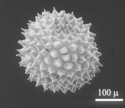
Figure 9.2.
A spiny microfossil found commonly in Doushantuo cherts and phosphatic rocks. Such fossils are thought to be the reproductive spores of eukaryotic organisms. Fossil is 250 microns in diameter. (Image courtesy of Shuhai Xiao)
The Maiohe fossils were preserved by rapid burial in fine-grained sediment. Bacteria normally decompose algal tissues soon after death, but at Maiohe they were prevented from completing this task by a veneer of clay that both excluded oxygen and adsorbed the enzymes that break cells apart. As sedimentary layers accumulated, biological remains were pressed like flowers between the pages of a book.
Leaves are commonly compressed in younger mudstones formed in lakes or the floodplains of rivers. In marine rocks, however, organic compressions are rare because burrowing animals irrigate and churn mud and silt layers. Rare, but not unknown. In fact, the most famous of all fossil deposits, the Middle Cambrian Burgess Shale, formed by the
compression of animal carcasses in carbon-rich mudstones.
4
Burgess fossils postdate Doushantuo deposition by only 50–85 million years; thus, the Doushantuo compressions are noteworthy for what they lack as well as what they include. One population of flanged tubes may record simple, sea anemone–like invertebrates (
plate 5b
), but nowhere do we find evidence of the anatomically and morphologically complex animals that are so conspicuous in Burgess and other Cambrian glimpses through the same preservational window. Evidently, much happened between Doushantuo and Burgess time.
I became involved in the study of Doushantuo fossils at the invitation of Professor Zhang Yun, a kind, cultured, and wonderfully insightful paleontologist at Beijing University. During the 1980s, Zhang collected Doushantuo samples from a phosphate mine near the Guizhou village of Weng’an. Several contained multicellular algae. In 1992, to my everlasting good fortune, he asked me to join him in collaborative research. By coincidence, a second Chinese friend, Yin Leiming of the Nanjing Institute of Geology and Palaeontology, invited me to visit China at about the same time. Yin was working on Doushantuo fossils in cherts from the Yangtze Gorges and Zhang was investigating assemblages from the phosphate mines of Guizhou, so I suggested that the three of us join forces to try to understand the full diversity of Doushantuo eukaryotes. Shuhai Xiao, who had completed his bachelor’s degree in Beijing under Zhang’s guidance, entered Harvard as a graduate student the same year, completing our team.
Doushantuo cherts and shales open two distinct and unusually clear windows on late Proterozoic life, but those Guizhou phosphates provide a third view, even better than the others. In this corner of the shallow Doushantuo seaway, biological remains that entered surface sediments were coated almost immediately by minute crystals of calcium phosphate minerals, preserving both overall morphology and cellular anatomy in remarkable three-dimensional detail. As a result, the Doushantuo phosphates preserve organisms seldom seen in rocks of any age.
Like cherts along the Yangtze River, the Guizhou phosphates are chock-full of highly ornamented eukaryotic microfossils; in some beds these fossils are so abundant that they literally form sandstones made of phosphatized cells. Other fossils are multicellular, and once again details of morphology show that most represent tissue-forming algae, not bacterial colonies. Particularly informative are small, crust-forming structures made of thick-walled cells arranged in rows that fan outward, like water as it gushes from a spring (
plate 5d
and
e
). Biologists call this anatomical organization a “cell fountain,” and it is especially common in red algae. Distinctive reproductive structures and anatomically distinct inner and outer tissues strengthen the ties between these fossils and a particular group of reds called the corallines. (Doushantuo fossils don’t display all of the features that collectively define coralline algae, but they exhibit enough of them to suggest that these small phosphatic fossils record an early way station in red algal evolution.) Other Guizhou fossils seem to lie halfway between the two major branches of living red algae, again suggesting that Doushantuo phosphates captured the diversity of red algae
in statu nascendi
—as it began to unfold.
Taken together, compressed and phosphatized Doushantuo fossils show that by the time large animals appeared in the oceans, multicellularity was already well established among the algae.
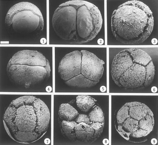
It is exciting to discover algae with cells preserved intact, but the crown jewels of Doushantuo are undoubtedly small balls, 400–500 microns in diameter, found in phosphates near the village of Weng’an (
figure 9.3
). The balls are uniform in size. Some contain a single cell wrapped in a thick furrowed coat, while others contain multiple cells surrounded by a thin membrane—cells in pairs, quartets, octads, and larger powers of two arranged in a geometric pattern determined by precisely oriented cell divisions. Parts of this division series were reported by Chinese paleontolgists in 1995 and interpreted as colonial green algae, but size, geometry, and envelope formation collectively make such an interpretation unlikely.
Shuhai Xiao discovered new and more informative populations that enabled him to recognize them as animals—specifically, animal eggs and embryos in the early stages of growth. Fossil embryos are rare in the geological record, but they do occur in Cambrian rocks. In fact, some beautiful Cambrian embryos described a year earlier by Stefan Bengtson of the Swedish Museum of Natural History and his Chinese colleague Zhang Yue were Shuhai’s inspiration to keep his eyes wide open when sorting Doushantuo fossils.