Pediatric Primary Care (180 page)

Figure 13
Impetigo. Spread of infection to top of nose from beneath the nose in an infant with impetigo.© Dr. P. Marazzi/Photo Researchers, Inc.
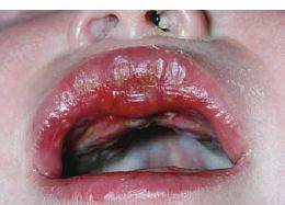
Figure 14
Infant with primary herpes gingivostomatitis.© Dr. P. Marazzi/Photo Researchers, Inc.
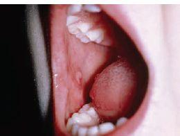
Figure 15
Erosion of the tongue in a child with hand-foot-and-mouth syndrome. © Dr. P. Marazzi/Photo Researchers, Inc.
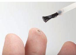
Figure 16
Common warts on child's finger. ©
leschnyhan/Fotolia.com
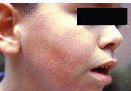
Figure 17
Slapped-cheek appearance of a child with parvovirus B19 infection (erythema infectiosum). Courtesy of CDC
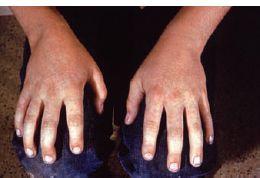
Figure 18
Lacy pink eruption over the palms in erythema infectiosum. Courtesy of CDC
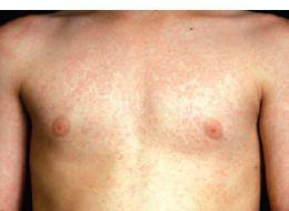
Figure 19
Lacy pink eruption of erythema infectiosum on chest and upper abdomen. Courtesy of Dr. Gary P. Williams, M. D.
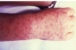
Figure 20
CDC-1962. RMSF, acral dusky oval purpuric lesions. Courtesy of CDC
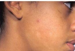
Figure 21
Pityriasis alba. In some atopic patients, subtle inflammation may result in poorly demarcated areas of hypopigmentation, known as pityriasis alba. Lesions are most prominent in darkly pigmented individuals.© Medical-on-Line/Alamy
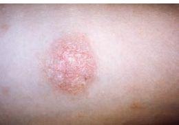
Figure 22
Pityriasis rosea. The large herald patch on the chest of this 10-year old girl shows central clearing, which mimics tinea corporis. Courtesy of Dr. Sellars/CDC
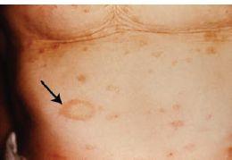
Figure 23
Pityriasis rosea. Numerous oval lesions on the chest of a Caucasian teenager. Courtesy of CDC
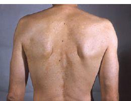
Figure 24
Tinea versicolor. The well-demarcated, scaly papules appear darker than surrounding skin on the back of a Caucasian adolescent. Courtesy of Dr. Lucille K. Georg/CDC
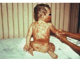
Figure 25
Roseaola/exanthema subitum. A generalized, pink, maculopapular rash suddenly appeared on this infant after 3 days of high fever. Courtesy of Arthur E. Kaye/CDC
