Ross & Wilson Anatomy and Physiology in Health and Illness (161 page)
Read Ross & Wilson Anatomy and Physiology in Health and Illness Online
Authors: Anne Waugh,Allison Grant
Tags: #Medical, #Nursing, #General, #Anatomy

Learning outcome
After studying this section you should be able to:
describe the structure of the bladder.
The urinary bladder is a reservoir for urine. It lies in the pelvic cavity and its size and position vary, depending on the volume of urine it contains. When distended, the bladder rises into the abdominal cavity.
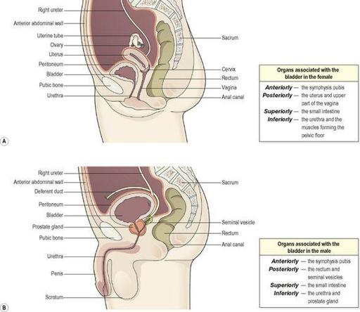
Organs associated with the bladder (
Fig. 13.20
)
Figure 13.20
The pelvic organs associated with the bladder and the urethra in: A.
The female.
B.
The male.
Structure (
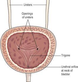
Fig. 13.21
)
The bladder is roughly pear shaped, but becomes more oval as it fills with urine. The posterior surface is the
base
. The bladder opens into the urethra at its lowest point, the
neck
.
Figure 13.21
Section of the bladder showing the trigone.
The peritoneum covers only the superior surface before it turns upwards as the parietal peritoneum, lining the anterior abdominal wall. Posteriorly it surrounds the uterus in the female and the rectum in the male.
The bladder wall is composed of three layers:
•
the outer layer of loose connective tissue, containing blood and lymphatic vessels and nerves, covered on the upper surface by the peritoneum
•
the middle layer, consisting of interlacing smooth muscle fibres and elastic tissue loosely arranged in three layers. This is called the
detrusor muscle
and when it contracts, it empties the bladder
•
the mucosa, composed of transitional epithelium (
p. 35
) that readily permits distension of the bladder as it fills with urine.
When the bladder is empty the inner lining is arranged in folds, or rugae, which gradually disappear as it fills. The bladder is distensible but when it contains 300 to 400 ml, awareness of the need to pass urine is felt. The total capacity is rarely more than about 600 ml.
The three orifices in the bladder wall form a triangle or
trigone
(
Fig. 13.21
). The upper two orifices on the posterior wall are the openings of the ureters. The lower orifice is the opening into the urethra. The
internal urethral sphincter
, a thickening of the urethral smooth muscle layer in the upper part of the urethra, controls outflow of urine from the bladder. This sphincter is not under voluntary control.
Urethra
Learning outcome
After studying this section you should be able to:
outline the structure and function of the urethra in males and females.
The urethra is a canal extending from the neck of the bladder to the exterior, at the external urethral orifice. It is longer in the male than in the female.
The male urethra is associated with the urinary and the reproductive systems, and is described in detail in
Chapter 18
.
The female urethra is approximately 4 cm long and 6 mm in diameter. It runs downwards and forwards behind the symphysis pubis and opens at the
external urethral orifice
just in front of the vagina. The external urethral orifice is guarded by the
external urethral sphincter
, which is under voluntary control.
The wall of the female urethra has two main layers: an outer muscle layer and an inner lining of mucosa, which is continuous with that of the bladder. The muscle layer has two parts, an inner layer of smooth muscle that is under autonomic nerve control, and an outer layer of striated muscle surrounding it. The striated muscle forms the external urethral sphincter and is under voluntary control. The mucosa is supported by loose fibroelastic connective tissue containing blood vessels and nerves. Proximally it consists of transitional epithelium while distally it is composed of stratified epithelium.
Micturition


