Ross & Wilson Anatomy and Physiology in Health and Illness (66 page)
Read Ross & Wilson Anatomy and Physiology in Health and Illness Online
Authors: Anne Waugh,Allison Grant
Tags: #Medical, #Nursing, #General, #Anatomy

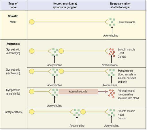
Figure 7.8
Neurotransmitters at synapses in the peripheral nervous system.
Somatic nerves carry impulses directly to the synapses at skeletal muscles, the
neuromuscular junctions
(
p. 411
) stimulating them to contract. In the autonomic nervous system (see
p. 167
), efferent impulses travel along two neurones (preganglionic and postganglionic) and across two synapses to the effector organs, e.g. smooth muscle and glands, in both the sympathetic and the parasympathetic divisions.
Nerves
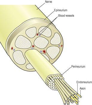
A nerve consists of numerous neurones collected into bundles (bundles of nerve fibres in the central nervous system are known as tracts). Each bundle has several coverings of protective connective tissue (
Fig. 7.9
).
•
Endoneurium
is a delicate tissue, surrounding each individual fibre, which is continuous with the septa that pass inwards from the perineurium.
•
Perineurium
is a smooth connective tissue, surrounding each
bundle
of fibres.
•
Epineurium
is the fibrous tissue which surrounds and encloses a number of bundles of nerve fibres. Most large nerves are covered by epineurium.
Figure 7.9
Transverse section of a peripheral nerve showing the protective coverings.
Sensory or afferent nerves
Sensory nerves carry information from the body to the spinal cord (
Fig. 7.1
). The impulses may then pass to the brain or to connector neurones of reflex arcs in the spinal cord (see
p. 158
).
Sensory receptors
Specialised endings of sensory neurones respond to different stimuli (changes) inside and outside the body.
Somatic, cutaneous or common senses
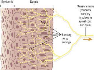
These originate in the skin. They are: pain, touch, heat and cold. Sensory nerve endings in the skin are fine branching filaments without myelin sheaths (
Fig. 7.10
). When stimulated, an impulse is generated and transmitted by the sensory nerves to the brain where the sensation is perceived.
Figure 7.10
Sensory nerve endings of the skin.
Proprioceptor senses
These originate in muscles and joints and contribute to the maintenance of balance and posture.
Special senses
These are sight, hearing, balance, smell and taste (see
Ch. 8
).
Autonomic afferent nerves
These originate in internal organs, glands and tissues, e.g. baroreceptors involved in the control of blood pressure (
Ch. 5
), chemoreceptors involved in the control of respiration (
Ch. 10
), and are associated with reflex regulation of involuntary activity and visceral pain.
Motor or efferent nerves
Motor nerves originate in the brain, spinal cord and autonomic ganglia. They transmit impulses to the effector organs: muscles and glands (
Fig. 7.1
). There are two types:
•
somatic nerves
– involved in voluntary and reflex skeletal muscle contraction
•
autonomic nerves
(sympathetic and parasympathetic) – involved in cardiac and smooth muscle contraction and glandular secretion.
Mixed nerves
In the spinal cord, sensory and motor nerves are arranged in separate groups, or
tracts
. Outside the spinal cord, when sensory and motor nerves are enclosed within the same sheath of connective tissue they are called
mixed nerves
.
Neuroglia
The neurones of the central nervous system are supported by four types of non-excitable
glial cells
that greatly outnumber the neurones (
Fig. 7.11
). Unlike nerve cells, which cannot divide, glial cells continue to replicate throughout life. They are
astrocytes, oligodendrocytes, ependymal cells
and
microglia
.
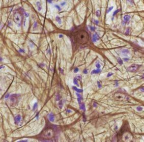
Figure 7.11
Neurones and glial cells.
A stained light micrograph of neurones (gold) and nuclei of the more numerous glial cells (blue).
Astrocytes
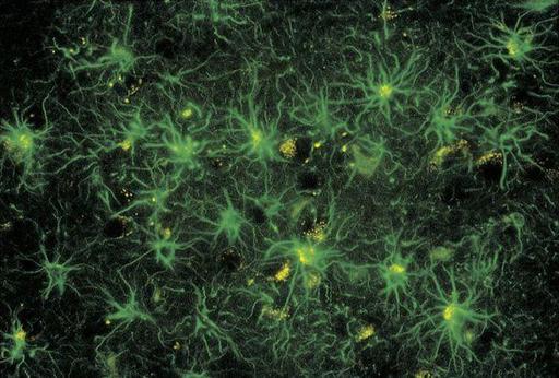
These cells form the main supporting tissue of the central nervous system. They are star shaped with fine branching processes and they lie in a mucopolysaccharide ground substance (
Fig. 7.12
). At the free ends of some of the processes are small swellings called
foot processes
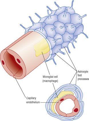
. Astrocytes are found in large numbers adjacent to blood vessels with their foot processes forming a sleeve round them. This means that the blood is separated from the neurones by the capillary wall and a layer of astrocyte foot processes which together constitute the
blood–brain barrier
(
Fig. 7.13
).
Figure 7.12
Star-shaped astrocytes in the cerebral cortex.
Figure 7.13
Blood–brain barrier. A.
Longitudinal section.
B.
Transverse section.
The blood–brain barrier is a selective barrier that protects the brain from potentially toxic substances and chemical variations in the blood, e.g. after a meal. Oxygen, carbon dioxide, alcohol, glucose and other lipid-soluble substances quickly cross the barrier into the brain. Some large molecules, drugs, inorganic ions and amino acids pass slowly from the blood to the brain.
Oligodendrocytes
These cells are smaller than astrocytes and are found in clusters round nerve cell bodies in grey matter; where they are thought to have a supportive function; adjacent to, and along the length of, myelinated nerve fibres. The oligodendrocytes form and maintain myelin, having the same functions as Schwann cells in peripheral nerves.
Ependymal cells
These cells form the epithelial lining of the ventricles of the brain and the central canal of the spinal cord. Those cells that form the choroid plexuses of the ventricles secrete cerebrospinal fluid.
Microglia
These cells may be derived from monocytes that migrate from the blood into the nervous system before birth. They are found mainly in the area of blood vessels. They enlarge and become phagocytic, removing microbes and damaged tissue, in areas of inflammation and cell destruction.
Response of nervous tissue to injury
Neurones reach maturity a few weeks after birth and cannot be replaced.
Damage to neurones can either lead to rapid necrosis with sudden acute functional failure, or to slow atrophy with gradually increasing dysfunction. These changes are associated with:
•
hypoxia and anoxia
•
nutritional deficiencies
•
poisons, e.g. organic lead
•
trauma
•
infections
•
ageing
•
hypoglycaemia.
Peripheral nerve regeneration (
Fig. 7.14
)
The axons of
peripheral nerves
can sometimes regenerate if the cell body remains intact. Distal to the damage, the axon and myelin sheath disintegrate and are removed by macrophages, but the Schwann cells survive and proliferate within the neurilemma. The live proximal part of the axon grows along the original track (about 1.5 mm per day), provided the two parts of neurilemma are correctly positioned and in close apposition (
Fig. 7.14A
). Restoration of function depends on the re-establishment of satisfactory connections with the effector organ. When the neurilemma is out of position or destroyed, the sprouting axons and Schwann cells form a tumour-like cluster (
traumatic neuroma
) producing severe pain, e.g. following some fractures and amputation of limbs (
Fig. 7.14B
).
