Ross & Wilson Anatomy and Physiology in Health and Illness (70 page)
Read Ross & Wilson Anatomy and Physiology in Health and Illness Online
Authors: Anne Waugh,Allison Grant
Tags: #Medical, #Nursing, #General, #Anatomy

•
sensory, which receive and decode sensory impulses enabling sensory perception
•
association, which are concerned with integration and processing of complex mental functions such as intelligence, memory, reasoning, judgement and emotions.
Figure 7.21
The cerebrum showing the main functional areas.
In general, motor impulses leave from the anterior part of each cerebral hemisphere while sensory impulses arrive at the posterior part, i.e. areas behind the central sulcus.
Motor areas of the cerebral cortex
The primary motor area
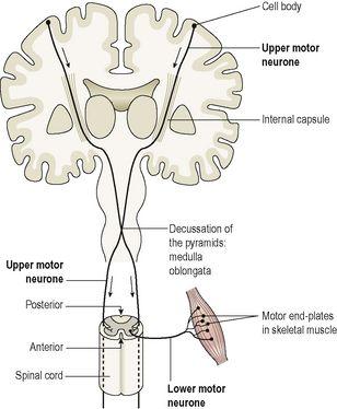
This lies in the frontal lobe immediately anterior to the central sulcus. The cell bodies are pyramid shaped (Betz’s cells) and they control skeletal muscle activity. There are two neurones involved in the pathway to skeletal muscle. The first, the
upper motor neurone,
descends from the motor cortex through the internal capsule to the medulla oblongata. Here it crosses to the opposite side and descends in the spinal cord. At the appropriate level in the spinal cord it synapses with a second neurone (the
lower motor neurone
), which leaves the spinal cord and travels to the target muscle. It terminates at the motor end plate of a muscle fibre (
Fig. 7.22
). This means that the motor area of the right hemisphere of the cerebrum controls voluntary muscle movement on the left side of the body and vice versa. Damage to either of these neurones may result in paralysis.
Figure 7.22
The motor nerve pathways:
upper and lower motor neurones.
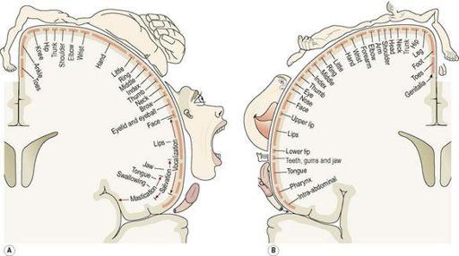
In the motor area of the cerebrum the body is represented upside down, i.e. the uppermost cells control the feet and those in the lowest part control the head, neck, face and fingers (
Fig. 7.23A
). The sizes of the areas of cortex representing different parts of the body are proportional to the complexity of movement of the body part, not to its size.
Figure 7.23A
shows that, in comparison with the trunk, the hand, foot, tongue and lips are represented by large cortical areas reflecting the greater degree of motor control associated with these areas.
Figure 7.23
A.
The motor homunculus showing how the body is represented in the motor area of the cerebrum.
B.
The sensory homunculus showing how the body is represented in the sensory area of the cerebrum.
(Both
A
and
B
are from Penfield W, Rasmussen T 1950 The cerebral cortex of man. Macmillan, New York. © 1950 Macmillan Publishing Co., renewed 1978 Theodore Rasmussen.)
Broca’s (motor speech) area
This is situated in the frontal lobe just above the lateral sulcus and controls the muscle movements needed for speech. It is dominant in the left hemisphere in right-handed people and vice versa.
Sensory areas of the cerebral cortex
The somatosensory area
This is the area immediately behind the central sulcus. Here sensations of pain, temperature, pressure and touch, awareness of muscular movement and the position of joints (proprioception) are perceived. The somatosensory area of the right hemisphere receives impulses from the left side of the body and vice versa. The size of the cortical areas representing different parts of the body (
Fig. 7.23B
) is proportional to the extent of sensory innervation, e.g. the large area for the face is consistent with the extensive sensory nerve supply by the three branches of the trigeminal nerves (5th cranial nerves).
The auditory (hearing) area
This lies immediately below the lateral sulcus within the temporal lobe. The nerve cells receive and interpret impulses transmitted from the inner ear by the cochlear (auditory) part of the vestibulocochlear nerves (8th cranial nerves).
The olfactory (smell) area
This lies deep within the temporal lobe where impulses from the nose, transmitted via the olfactory nerves (1st cranial nerves), are received and interpreted.
The taste area
This lies just above the lateral sulcus in the deep layers of the somatosensory area. Here, impulses from sensory receptors in taste buds are received and perceived as taste.
The visual area
This lies behind the parieto-occipital sulcus and includes the greater part of the occipital lobe. The optic nerves (2nd cranial nerves) pass from the eye to this area, which receives and interprets the impulses as visual impressions.
Association areas
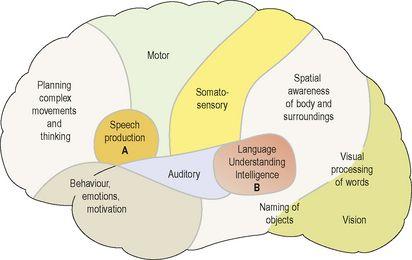
These are connected to each other and other areas of the cerebral cortex by association tracts and some are outlined below. They receive, coordinate and interpret impulses from the sensory and motor cortices permitting higher cognitive abilities and, although
Figure 7.24
depicts some of the areas involved, their functions are much more complex.
Figure 7.24
Areas of the cerebral cortex involved in higher mental functions. A.
Broca’s area.
B.
Wernicke’s area.
The premotor area
This lies in the frontal lobe immediately anterior to the motor area. The neurones here coordinate movement initiated by the primary motor cortex, ensuring that learned patterns of movement can be repeated. For example, in tying a shoelace or writing, many muscles contract but the movements must be coordinated and carried out in a particular sequence. Such a pattern of movement, when established, is described as
manual dexterity
.
The prefrontal area
This extends anteriorly from the premotor area to include the remainder of the frontal lobe. It is a large area and is more highly developed in humans than in other animals. Intellectual functions controlled here include perception and comprehension of the passage of time, the ability to anticipate consequences of events and the normal management of emotions.
Wernicke’s (sensory speech) area
This is situated in the temporal lobe adjacent to the parieto-occipitotemporal area. It is here that the spoken word is perceived, and comprehension and intelligence are based. Understanding language is central to higher mental functions as they are language based. This area is dominant in the left hemisphere in right-handed people and vice versa.
The parieto-occipitotemporal area
This lies behind the somatosensory area and includes most of the parietal lobe. Its functions are thought to include spatial awareness, interpreting written language and the ability to name objects (
Fig. 7.24
). It has been suggested that objects can be recognised by touch alone because of the knowledge from past experience (memory) retained in this area.
Diencephalon (see
Fig. 7.18
)
This part of the brain connects the cerebrum and the midbrain. It consists of several structures situated around the third ventricle, the main ones being the thalamus and hypothalamus, which are considered here. The pineal gland (
p. 219
) and the optic chiasma (
p. 194
) are situated there.
Thalamus
This consists of two masses of grey and white matter situated within the cerebral hemispheres just below the corpus callosum, one on each side of the third ventricle (
Fig. 7.18
). Sensory receptors in the skin and viscera send information about touch, pain and temperature, and input from the special sense organs travels to the thalamus where there is recognition, although only in a basic form, as refined perception also involves other parts of the brain. It is thought to be involved in the processing of some emotions and complex reflexes. The thalamus relays and redistributes impulses from most parts of the brain to the cerebral cortex.
Hypothalamus
The hypothalamus is a small but important structure which weighs around 7 g and consists of a number of nuclei. It is situated below and in front of the thalamus, immediately above the
pituitary gland
. The hypothalamus is linked to the posterior lobe of the pituitary gland by nerve fibres and to the anterior lobe by a complex system of blood vessels. Through these connections, the hypothalamus controls the output of hormones from both lobes of the pituitary gland (see
p. 209
).
Other functions of the hypothalamus include control of:
•
the autonomic nervous system (
p. 167
)
•
appetite and satiety
•
thirst and water balance
•
body temperature (
p. 358
)
•
emotional reactions, e.g. pleasure, fear, rage
•
sexual behaviour and child rearing
•
sleeping and waking cycles.
Brain stem (
Fig. 7.18
)
Midbrain
The midbrain is the area of the brain situated around the cerebral aqueduct between the cerebrum above and the
pons
below. It consists of nuclei and nerve fibres (tracts), which connect the cerebrum with lower parts of the brain and with the spinal cord. The nuclei act as relay stations for the ascending and descending nerve fibres.
Pons


