Ross & Wilson Anatomy and Physiology in Health and Illness (36 page)
Read Ross & Wilson Anatomy and Physiology in Health and Illness Online
Authors: Anne Waugh,Allison Grant
Tags: #Medical, #Nursing, #General, #Anatomy

explain the relationship between the different types of blood vessel
indicate the main factors controlling blood vessel diameter
explain the mechanisms by which exchange of nutrients, gases and wastes occurs between the blood and the tissues.
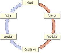
Blood vessels vary in structure, size and function, and there are several types: arteries, arterioles, capillaries, venules and veins (
Fig. 5.2
).
Figure 5.2
The relationship between the heart and the different types of blood vessel.
Arteries and arterioles
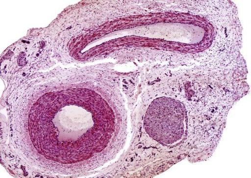
These are the blood vessels that transport blood away from the heart. They vary considerably in size and their walls consist of three layers of tissue (
Fig. 5.3
):
•
tunica adventitia
or outer layer of fibrous tissue
•
tunica media
or middle layer of smooth muscle and elastic tissue
•
tunica intima
or inner lining of squamous epithelium called
endothelium
.
Figure 5.3
Light micrograph of an artery, a vein and an associated nerve.
The amount of muscular and elastic tissue varies in the arteries depending upon their size and function. In the large arteries, sometimes called elastic arteries, the tunica media consists of more elastic tissue and less smooth muscle. This allows the vessel wall to stretch, absorbing the pressure wave generated by the heart as it beats. These proportions gradually change as the arteries branch many times and become smaller until in the
arterioles
(the smallest arteries) the tunica media consists almost entirely of smooth muscle. This enables their diameter to be precisely controlled, which regulates the pressure within them. Systemic blood pressure is mainly determined by the resistance these tiny arteries offer to blood flow, and for this reason they are called
resistance vessels
.
Arteries have thicker walls than veins to withstand the high pressure of arterial blood.
Anastomoses and end-arteries
Anastomoses
are arteries that form a link between main arteries supplying an area, e.g. the arterial supply to the palms of the hand (
p. 98
) and soles of the feet, the brain, the joints and, to a limited extent, the heart muscle. If one artery supplying the area is occluded, anastomotic arteries provide a
collateral circulation
. This is most likely to provide an adequate blood supply when the occlusion occurs gradually, giving the anastomotic arteries time to dilate.
End
-
arteries
are the arteries with no anastomoses or those beyond the most distal anastomosis, e.g. the branches from the circulus arteriosus (circle of Willis) in the brain or the central artery to the retina of the eye. When an end-artery is occluded the tissues it supplies die because there is no alternative blood supply.
Capillaries and sinusoids
The smallest arterioles break up into a number of minute vessels called
capillaries
. Capillary walls consist of a single layer of endothelial cells sitting on a very thin basement membrane, through which water and other small molecules can pass. Blood cells and large molecules such as plasma proteins do not normally pass through capillary walls. The capillaries form a vast network of tiny vessels that link the smallest arterioles to the smallest venules. Their diameter is approximately that of an erythrocyte (7 μm). The capillary bed is the site of exchange of substances between the blood and the tissue fluid, which bathes the body cells.
Entry to capillary beds is guarded by rings of smooth muscle (
precapillary sphincters
) that direct blood flow. Hypoxia (low levels of oxygen in the tissues), or high levels of tissue wastes, indicating high levels of activity, dilate the sphincters and increase blood flow through the affected beds.
In certain places, including the liver and bone marrow, the capillaries are significantly wider and leakier than normal. These capillaries are called
sinusoids
and because their walls are incomplete and their lumen is much larger than usual, the blood flows through them more slowly under less pressure and can come directly into contact with the cells outside the sinusoid wall. This allows much faster exchange of substances between the blood and the tissues, useful, for example, in the liver, which regulates the composition of blood arriving from the gastrointestinal tract.
Veins and venules
Veins are blood vessels that return blood at low pressure to the heart. The walls of the veins are thinner than those of arteries but have the same three layers of tissue (
Fig. 5.3
). They are thinner because there is less muscle and elastic tissue in the tunica media, because veins carry blood at a lower pressure than arteries. When cut, the veins collapse while the thicker-walled arteries remain open.
When an artery is cut blood spurts at high pressure while a slower, steady flow of blood escapes from a vein.
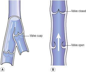
Some veins possess
valves
, which prevent backflow of blood, ensuring that it flows towards the heart (
Fig. 5.4
). They are formed by a fold of tunica intima and strengthened by connective tissue. The cusps are
semilunar
in shape with the concavity towards the heart. Valves are abundant in the veins of the limbs, especially the lower limbs where blood must travel a considerable distance against gravity when the individual is standing. They are absent in very small and very large veins in the thorax and abdomen. Valves are assisted in maintaining one-way flow by skeletal muscles surrounding the veins (
p. 86
).
Figure 5.4
Interior of a vein: A.
The valves and cusps.
B.
The direction of blood flow through a valve.
The smallest veins are called
venules
.
Veins are called
capacitance vessels
because they are distensible, and therefore have the capacity to hold a large proportion of the body’s blood. At any one time, about two-thirds of the body’s blood is in the venous system. This allows the vascular system to absorb (to an extent) sudden changes in blood volume, such as in haemorrhage; the veins can recoil, helping to prevent a sudden fall in blood pressure.


