Ross & Wilson Anatomy and Physiology in Health and Illness (38 page)
Read Ross & Wilson Anatomy and Physiology in Health and Illness Online
Authors: Anne Waugh,Allison Grant
Tags: #Medical, #Nursing, #General, #Anatomy

Heart
Learning outcomes
After studying this section, you should be able to:
describe the structure of the heart and its position within the thorax
trace the circulation of the blood through the heart and the blood vessels of the body
outline the conducting system of the heart
relate the electrical activity of the cardiac conduction system to the cardiac cycle
describe the main factors determining heart rate and cardiac output.
The heart is a roughly cone-shaped hollow muscular organ. It is about 10 cm long and is about the size of the owner’s fist. It weighs about 225 g in women and is heavier in men (about 310 g).
Position
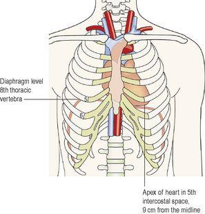
The heart lies in the thoracic cavity (
Fig. 5.9
) in the mediastinum (the space between the lungs). It lies obliquely, a little more to the left than the right, and presents a base above, and an
apex
below. The apex is about 9 cm to the left of the midline at the level of the 5th intercostal space, i.e. a little below the nipple and slightly nearer the midline. The base extends to the level of the 2nd rib.
Figure 5.9
Position of the heart in the thorax.
Organs associated with the heart (
Fig. 5.10
)
Inferiorly
– the apex rests on the central tendon of the diaphragm
Superiorly
– the great blood vessels, i.e. the aorta, superior vena cava, pulmonary artery and pulmonary veins
Posteriorly
– the oesophagus, trachea, left and right bronchus, descending aorta, inferior vena cava and thoracic vertebrae
Laterally
– the lungs – the left lung overlaps the left side of the heart
Anteriorly
– the sternum, ribs and intercostal muscles.
Figure 5.10
Organs associated with the heart.
Structure
The heart wall
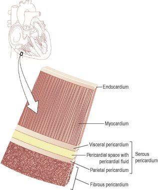
The heart wall is composed of three layers of tissue (
Fig. 5.11
): pericardium, myocardium and endocardium.
Figure 5.11
Layers of the heart wall.
Pericardium
The pericardium is the outermost layer and is made up of two sacs. The outer sac consists of fibrous tissue and the inner of a continuous double layer of serous membrane.
The outer fibrous sac is continuous with the tunica adventitia of the great blood vessels above and is adherent to the diaphragm below. Its inelastic, fibrous nature prevents overdistension of the heart.
The outer layer of the serous membrane, the
parietal pericardium
, lines the fibrous sac. The inner layer, the
visceral pericardium
, or epicardium, which is continuous with the parietal pericardium, is adherent to the heart muscle. A similar arrangement of a double membrane forming a closed space is seen also with the pleura, the membrane enclosing the lungs (see
Fig. 10.15, p. 243
).


