Ross & Wilson Anatomy and Physiology in Health and Illness (48 page)
Read Ross & Wilson Anatomy and Physiology in Health and Illness Online
Authors: Anne Waugh,Allison Grant
Tags: #Medical, #Nursing, #General, #Anatomy

Paired testicular, ovarian, renal and adrenal veins join the inferior vena cava.
Blood from the remaining organs in the abdominal cavity passes through the liver via the
portal circulation
before entering the inferior vena cava (
Fig. 5.44
).
Portal circulation
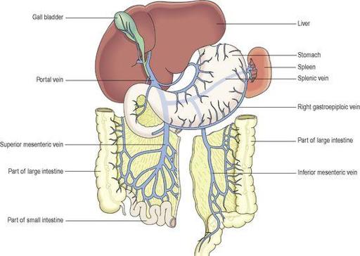
In all parts of circulation described so far, venous blood passes from the tissues to the heart by the most direct route through only one capillary bed. In the portal circulation, venous blood passes from the capillary beds of the abdominal part of the digestive system, the spleen and pancreas to the liver. It then passes through a second capillary bed, the hepatic sinusoids, in the liver before entering the general circulation via the inferior vena cava. In this way, blood with a high concentration of nutrients, absorbed from the stomach and intestines, goes to the liver first. In the liver certain modifications take place, including the regulation of blood nutrient levels.
Portal vein
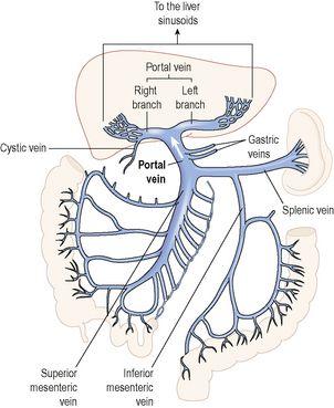
This is formed by the union of several veins (
Figs 5.46
and
5.47
), each of which drains blood from the area supplied by the corresponding artery:
•
The
splenic vein
drains blood from the spleen, the pancreas and part of the stomach.
•
The
inferior mesenteric vein
returns the venous blood from the rectum, pelvic and descending colon of the large intestine. It joins the splenic vein.
•
The
superior mesenteric vein
returns venous blood from the small intestine and the proximal parts of the large intestine, i.e. the caecum, ascending and transverse colon. It unites with the splenic vein to form the portal vein.
•
The
gastric veins
drain blood from the stomach and the distal end of the oesophagus, then join the portal vein.
•
The
cystic vein,
which drains venous blood from the gall bladder, joins the portal vein.
Figure 5.46
Venous drainage from the abdominal organs, and the formation of the portal vein.
Figure 5.47
The portal vein – origin and termination.
Hepatic veins
These are very short veins that leave the posterior surface of the liver and, almost immediately, enter the inferior vena cava.
Circulation to the pelvis and lower limb
Arterial supply
Common iliac arteries
The right and left common iliac arteries are formed when the abdominal aorta divides at the level of the 4th lumbar vertebra (
Fig. 5.28
). In front of the sacroiliac joint each divides into the internal and the external iliac arteries.
The
internal iliac artery
runs medially to supply the organs within the pelvic cavity. In the female, one of the largest branches is the uterine artery, which provides the main arterial blood supply to the reproductive organs.
The
external iliac artery
runs obliquely downwards and passes behind the inguinal ligament into the thigh where it becomes the femoral artery.
The
femoral artery
(
Fig. 5.48
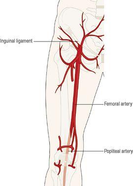
) begins at the midpoint of the inguinal ligament and extends downwards in front of the thigh; it then turns medially and eventually passes round the medial aspect of the femur to enter the popliteal space where it becomes the popliteal artery. It supplies blood to the structures of the thigh and some superficial pelvic and inguinal structures.
Figure 5.48
The femoral artery and its main branches.
The
popliteal artery
(
Fig. 5.49
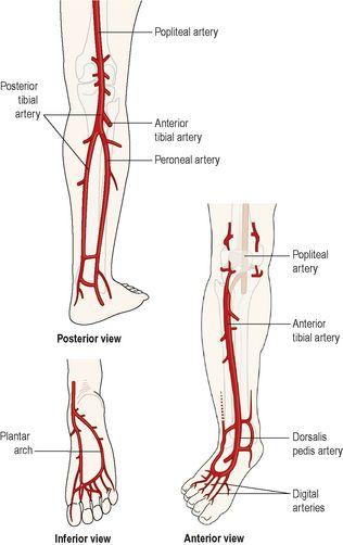
) passes through the popliteal fossa behind the knee, where the pulse can be felt. It supplies the structures in this area, including the knee joint. At the lower border of the popliteal fossa it divides into the anterior and posterior tibial arteries.
Figure 5.49
The right popliteal artery and its main branches.
The
anterior tibial artery
(
Fig. 5.49
) passes forwards between the tibia and fibula and supplies the structures in the front of the leg. It lies on the tibia, runs in front of the ankle joint and continues over the dorsum (top) of the foot as the
dorsalis pedis artery
.
The
dorsalis pedis artery
is a continuation of the anterior tibial artery and passes over the dorsum of the foot, where the pulse can be felt, supplying arterial blood to the structures in this area. It ends by passing between the first and second metatarsal bones into the sole of the foot where it contributes to the formation of the plantar arch.
The
posterior tibial artery
(
Fig. 5.49
) runs downwards and medially on the back of the leg. Near its origin it gives off a large branch called the
peroneal artery
, which supplies the lateral aspect of the leg. In the lower part it becomes superficial and passes medial to the ankle joint to reach the sole of the foot, where it continues as the
plantar artery
.
The
plantar artery
supplies the structures in the sole of the foot. This artery, its branches and the dorsalis pedis artery form the
plantar arch
from which the digital branches arise to supply the toes.
Venous return
There are both deep and superficial veins in the lower limb (
Fig. 5.29
). Blood entering the superficial veins passes to the deep veins through
communicating veins
. Movement of blood towards the heart is partly dependent on contraction of skeletal muscles. Backward flow is prevented by a large number of valves. Superficial veins receive less support from surrounding tissues than deep veins.
Deep veins
The deep veins accompany the arteries and their branches and have the same names. They are the:
•
femoral vein
, which ascends in the thigh to the level of the inguinal ligament, where it becomes the external iliac vein
•
external iliac vein
, the continuation of the femoral vein where it enters the pelvis lying close to the femoral artery. It passes along the brim of the pelvis, and at the level of the sacroiliac joint it is joined by the
internal iliac vein
to form the
common iliac vein
•
internal iliac vein
, which receives tributaries from several veins draining the organs of the pelvic cavity
•
two common iliac veins
, which begin at the level of the sacroiliac joints. They ascend obliquely and end a little to the right of the body of the 5th lumbar vertebra by uniting to form the
inferior vena cava
.
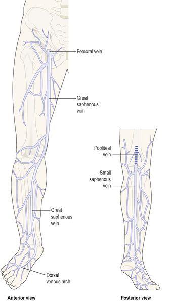
Superficial veins (
Fig. 5.50
)
The two main superficial veins draining blood from the lower limbs are the small and the great saphenous veins.
Figure 5.50
Superficial veins of the leg.
The
small saphenous vein
begins behind the ankle joint where many small veins which drain the dorsum of the foot join together. It ascends superficially along the back of the leg and in the popliteal space it joins the
popliteal vein
– a deep vein.
The
great saphenous vein
is the longest vein in the body. It begins at the medial half of the dorsum of the foot and runs upwards, crossing the medial aspect of the tibia and up the inner side of the thigh. Just below the inguinal ligament it joins the
femoral vein
.
Many
communicating veins
join the superficial veins, and the superficial and deep veins of the lower limb.
Summary of the main blood vessels (
Fig. 5.51
)
