Ross & Wilson Anatomy and Physiology in Health and Illness (45 page)
Read Ross & Wilson Anatomy and Physiology in Health and Illness Online
Authors: Anne Waugh,Allison Grant
Tags: #Medical, #Nursing, #General, #Anatomy

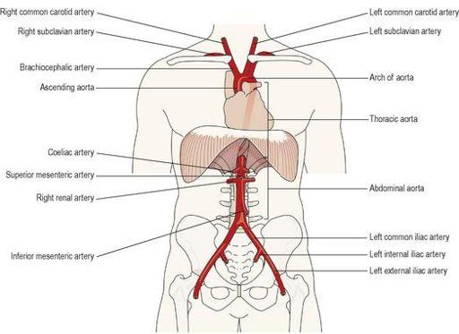
Figure 5.28
The aorta and the main arteries of the limbs.
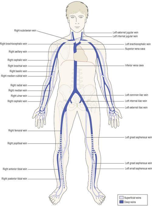
Figure 5.29
The venae cavae and the main veins of the limbs.
Pulmonary circulation
This is the circulation of blood from the right ventricle of the heart to the lungs and back to the left atrium. In the lungs, carbon dioxide is excreted and oxygen is absorbed.
The pulmonary artery
or trunk, carrying
deoxygenated blood
, leaves the upper part of the right ventricle of the heart. It passes upwards and divides into left and right pulmonary arteries at the level of the 5th thoracic vertebra.
The left pulmonary artery
runs to the root of the left lung where it divides into two branches, one passing into each lobe.
The right pulmonary artery
passes to the root of the right lung and divides into two branches. The larger branch carries blood to the middle and lower lobes, and the smaller branch to the upper lobe.
Within the lung these arteries divide and subdivide into smaller arteries, arterioles and capillaries. The exchange of gases takes place between capillary blood and air in the alveoli of the lungs (
p. 250
). In each lung the capillaries containing oxygenated blood join up and eventually form two pulmonary veins.
Two pulmonary veins
leave each lung, returning oxygenated blood to the left atrium of the heart. During atrial systole this blood is pumped into the left ventricle, and during ventricular systole it is forced into the aorta, the first artery of the general circulation.
Systemic or general circulation
The blood pumped out from the left ventricle is carried by the branches of the
aorta
around the body and returns to the right atrium of the heart by the
superior
and
inferior venae cavae
.
Figure 5.28
shows the general positions of the aorta and the main arteries of the limbs.
Figure 5.29
provides an overview of the venae cavae and the veins of the limbs.
The circulation of blood to the different parts of the body will be described in the order in which their arteries branch off the aorta.
Aorta
The aorta (
Fig. 5.30
) begins at the upper part of the left ventricle and, after passing upwards for a short way, it arches backwards and to the left. It then descends behind the heart through the thoracic cavity a little to the left of the thoracic vertebrae. At the level of the 12th thoracic vertebra it passes behind the diaphragm then downwards in the abdominal cavity to the level of the 4th lumbar vertebra, where it divides into the
right
and
left common iliac arteries
.
Figure 5.30
The aorta and its main branches.
Throughout its length the aorta gives off numerous branches. Some of the branches are
paired
, i.e. there is a right and left branch of the same name, for instance, the right and left renal arteries supplying the kidneys, and some are single or
unpaired
, e.g. the coeliac artery.
The aorta will be described here according to its location:
•
thoracic aorta (see below)
•
abdominal aorta (
p. 100
).
Thoracic aorta
This part of the aorta lies above the diaphragm and is described in three parts:
•
ascending aorta
•
arch of the aorta
•
descending aorta in the thorax (
p. 99
).
Ascending aorta
This is the short section of the aorta that rises from the heart. It is about 5 cm long and lies behind the sternum.
The right and left coronary arteries
are its only branches and they arise from the aorta just above the level of the aortic valve (
Fig. 5.16
). These important arteries supply the myocardium.
Arch of the aorta
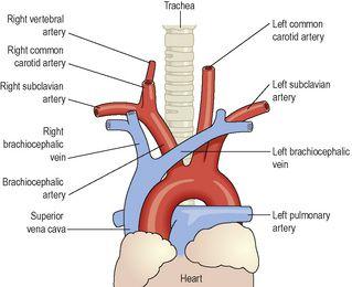
The arch of the aorta is a continuation of the ascending aorta. It begins behind the manubrium of the sternum and runs upwards, backwards and to the left in front of the trachea. It then passes downwards to the left of the trachea and is continuous with the descending aorta.
Three branches are given off from its upper aspect (
Fig. 5.31
):
•
brachiocephalic artery or trunk
•
left common carotid artery
•
left subclavian artery.
Figure 5.31
The arch of the aorta and its branches.
The brachiocephalic artery
is about 4 to 5 cm long and passes obliquely upwards, backwards and to the right. At the level of the sternoclavicular joint it divides into the
right common carotid artery
and the
right subclavian artery
.
Circulation of blood to the head and neck
Arterial supply
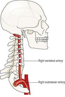
The paired arteries supplying the head and neck are the common carotid arteries and the vertebral arteries (
Figs 5.32
,
5.34
).
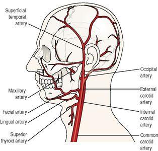
Figure 5.32
Main arteries of the left side of the head and neck.
Figure 5.34
The right vertebral artery.
Carotid arteries
The
right common carotid artery
is a branch of the brachiocephalic artery. The
left common carotid artery
arises directly from the arch of the aorta. They pass upwards on either side of the neck and have the same distribution on each side. The common carotid arteries are embedded in fascia, called the
carotid sheath
. At the level of the upper border of the thyroid cartilage each divides into an
internal carotid artery
and an
external carotid artery
.
The
carotid sinuses
are slight dilations at the point of division (bifurcation) of the common carotid arteries into their internal and external branches. The walls of the sinuses are thin and contain numerous nerve endings of the glossopharyngeal nerves. These nerve endings, or
baroreceptors
, are stimulated by changes in blood pressure in the carotid sinuses. The resultant nerve impulses initiate reflex adjustments of blood pressure through the vasomotor centre in the medulla oblongata (
p. 89
).
The
carotid bodies
are two small groups of specialised cells, called
chemoreceptors
, one lying in close association with each common carotid artery at its bifurcation. They are supplied by the glossopharyngeal nerves and their cells are stimulated by changes in the carbon dioxide and oxygen content of blood. The resultant nerve impulses initiate reflex adjustments of respiration through the respiratory centre in the medulla oblongata.
External carotid artery
(
Fig. 5.32
)This artery supplies the superficial tissues of the head and neck, via a number of branches:
•
The
superior thyroid artery
supplies the thyroid gland and adjacent muscles.
•
The
lingual artery
supplies the tongue, the lining membrane of the mouth, the structures in the floor of the mouth, the tonsil and the epiglottis.
•
The
facial artery
passes outwards over the mandible just in front of the angle of the jaw and supplies the muscles of facial expression and structures in the mouth. The pulse can be felt where the artery crosses the jaw bone.
•
The
occipital artery
supplies the posterior part of the scalp.
•
The
temporal artery
passes upwards over the zygomatic process in front of the ear and supplies the frontal, temporal and parietal parts of the scalp. The pulse can be felt in front of the upper part of the ear.
