Ross & Wilson Anatomy and Physiology in Health and Illness (75 page)
Read Ross & Wilson Anatomy and Physiology in Health and Illness Online
Authors: Anne Waugh,Allison Grant
Tags: #Medical, #Nursing, #General, #Anatomy

Figure 7.35
The brachial plexus.
Anterior view. Ant = anterior, Post = posterior.
The branches of the brachial plexus supply the skin and muscles of the upper limbs and some of the chest muscles. Five large nerves and a number of smaller ones emerge from this plexus, each with a contribution from more than one nerve root, containing sensory, motor and autonomic fibres:
•
axillary (circumflex) nerve: C5, 6
•
radial nerve: C5, 6, 7, 8, T1
•
musculocutaneous nerve: C5, 6, 7
•
median nerve: C5, 6, 7, 8, T1
•
ulnar nerve: C7, 8, T1
•
medial cutaneous nerve: C8, T1.
The axillary (circumflex) nerve
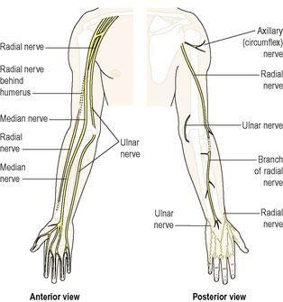
winds round the humerus at the level of the surgical neck. It then breaks up into minute branches to supply the deltoid muscle, shoulder joint and overlying skin.
The radial nerve
is the largest branch of the brachial plexus. It supplies the triceps muscle behind the humerus, crosses in front of the elbow joint then winds round to the back of the forearm to supply extensors of the wrist and finger joints. It continues into the back of the hand to supply the skin of the thumb, the first two fingers and the lateral half of the third finger.
The musculocutaneous nerve
passes downwards to the lateral aspect of the forearm. It supplies the muscles of the upper arm and the skin of the forearm.
The median nerve passes down the midline of the arm in close association with the brachial artery. It passes in front of the elbow joint then down to supply the muscles of the front of the forearm. It continues into the hand where it supplies small muscles and the skin of the front of the thumb, the first two fingers and the lateral half of the third finger. It gives off no branches above the elbow.
The ulnar nerve
descends through the upper arm lying medial to the brachial artery. It passes behind the medial epicondyle of the humerus to supply the muscles on the ulnar aspect of the forearm. It continues downwards to supply the muscles in the palm of the hand and the skin of the whole of the little finger and the medial half of the third finger. It gives off no branches above the elbow.
The main nerves of the arm are presented in
Figure 7.36
. The distribution and origins of the cutaneous sensory nerves of the arm, i.e. the dermatomes, are shown in
Figure 7.37
.
Figure 7.36
The main nerves of the arm.
Lumbar plexus (
Figs 7.38
,
7.40
and
7.41
)
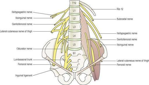
The lumbar plexus is formed by the anterior rami of the first three and part of the 4th lumbar nerves. The plexus is situated in front of the transverse processes of the lumbar vertebrae and behind the psoas muscle. The main branches, and their nerve roots are:
•
iliohypogastric nerve: L1
•
ilioinguinal nerve: L1
•
genitofemoral: L1, 2
•
lateral cutaneous nerve of thigh: L2, 3
•
femoral nerve: L2, 3, 4
•
obturator nerve: L2, 3, 4
•
lumbosacral trunk: L4, (5).
Figure 7.38
The lumbar plexus.
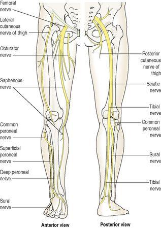
Figure 7.40
The main nerves of the leg.
The iliohypogastric
,
ilioinguinal
and
genitofemoral nerves
supply muscles and the skin in the area of the lower abdomen, upper and medial aspects of the thigh and the inguinal region.
The lateral cutaneous nerve of the thigh
supplies the skin of the lateral aspect of the thigh including part of the anterior and posterior surfaces.
The femoral nerve
is one of the larger branches. It passes behind the inguinal ligament to enter the thigh in close association with the femoral artery. It divides into cutaneous and muscular branches to supply the skin and the muscles of the front of the thigh. One branch, the
saphenous nerve
, supplies the medial aspect of the leg, ankle and foot.
The obturator nerve
supplies the adductor muscles of the thigh and skin of the medial aspect of the thigh. It ends just above the level of the knee joint.
The lumbosacral trunk
descends into the pelvis and makes a contribution to the sacral plexus.
Sacral plexus (
Figs 7.39
,
7.40
and
7.41
)
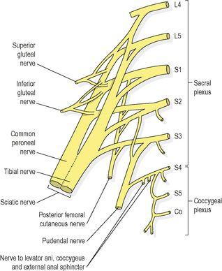
The sacral plexus is formed by the anterior rami of the lumbosacral trunk and the 1st, 2nd and 3rd sacral nerves. The lumbosacral trunk is formed by the 5th and part of the 4th lumbar nerves. It lies in the posterior wall of the pelvic cavity.
Figure 7.39
Sacral and coccygeal plexuses.
The sacral plexus divides into a number of branches, supplying the muscles and skin of the pelvic floor, muscles around the hip joint and the pelvic organs. In addition to these it provides the sciatic nerve, which contains fibres from L4 and 5 and S1–3.
The sciatic nerve
is the largest nerve in the body. It is about 2 cm wide at its origin. It passes through the greater sciatic foramen into the buttock then descends through the posterior aspect of the thigh supplying the hamstring muscles. At the level of the middle of the femur it divides to form the
tibial
and the
common peroneal nerves
.
The tibial nerve
descends through the popliteal fossa to the posterior aspect of the leg where it supplies muscles and skin. It passes under the medial malleolus to supply muscles and skin of the sole of the foot and toes. One of the main branches is the
sural nerve
, which supplies the tissues in the area of the heel, the lateral aspect of the ankle and a part of the dorsum of the foot.
The common peroneal nerve
descends obliquely along the lateral aspect of the popliteal fossa, winds round the neck of the fibula into the front of the leg where it divides into the
deep peroneal
(anterior tibial) and the
superficial peroneal
(musculocutaneous) nerves. These nerves supply the skin and muscles of the anterior aspect of the leg and the dorsum of the foot and toes.
The pudendal nerve
(S2, 3, 4) – the perineal branch supplies the external anal sphincter, the external urethral sphincter and adjacent skin.
Figures 7.40
and
7.41
show the main nerves of the leg, the dermatomes and the origins of the main nerves.
Coccygeal plexus (
Fig. 7.39
)
The coccygeal plexus
is a very small plexus formed by part of the 4th and 5th sacral and the coccygeal nerves. The nerves from this plexus supply the skin around the coccyx and anal area.
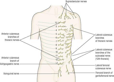
Thoracic nerves
The thoracic nerves
do not
intermingle to form plexuses. There are 12 pairs and the first 11 are the
intercostal nerves
. They pass between the ribs supplying them, the intercostal muscles and overlying skin. The 12th pair comprise the
subcostal nerves
. The 7th to the 12th thoracic nerves also supply the muscles and the skin of the posterior and anterior abdominal walls (
Fig. 7.42
).
Figure 7.42
Segmental distribution of the thoracic cutaneous nerves.
Cranial nerves (
Fig. 7.43
)
There are 12 pairs of cranial nerves originating from nuclei in the inferior surface of the brain, some sensory, some motor and some mixed. Their names generally suggest their distribution or function and they are numbered using Roman numerals according to the order they connect to the brain, starting anteriorly. They are:
