Ross & Wilson Anatomy and Physiology in Health and Illness (85 page)
Read Ross & Wilson Anatomy and Physiology in Health and Illness Online
Authors: Anne Waugh,Allison Grant
Tags: #Medical, #Nursing, #General, #Anatomy

8.3
The visual pathway
197
8.4
How the brain interprets odours
200
The special senses of hearing, sight, smell and taste all have specialised sensory receptors (nerve endings) outside the brain. These are found in the ears, eyes, nose and mouth. In the brain the incoming nerve impulses undergo complex processes of integration and coordination that result in perception of sensory information and a variety of responses inside and outside the body. Up to 80% of what we perceive comes from sensory stimuli. The first sections of this chapter explore the special senses, while the later ones consider problems that arise when disorders occur in the structures involved in hearing and vision.
Hearing and the ear
Learning outcomes
After studying this section you should be able to:
describe the structure of the outer, middle and inner parts of the ear
explain the physiology of hearing.
The ear is the organ of hearing and is also involved in balance. It is supplied by the 8th cranial nerve, i.e. the
cochlear part
of the
vestibulocochlear
nerve, which is stimulated by vibrations caused by sound waves.
With the exception of the auricle (pinna), the structures that form the ear are encased within the petrous portion of the temporal bone.
Structure
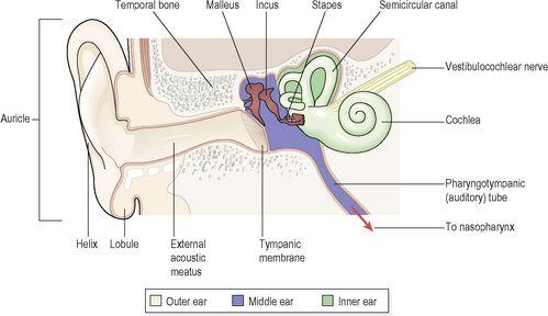
The ear is divided into three distinct parts (
Fig. 8.1
):
•
outer ear
•
middle ear (tympanic cavity)
•
inner ear.
Figure 8.1
The parts of the ear.
The outer ear collects the sound waves and directs them to the middle ear, which in turn transfers them to the inner ear, where they are converted to nerve impulses and transmitted to the hearing area in the cerebral cortex.
Outer ear
The outer ear consists of the auricle (pinna) and the external acoustic meatus (auditory canal).
The auricle (pinna)
The auricle is the visible part of the ear that projects from the side of the head. It is composed of fibroelastic cartilage covered with skin. It is deeply grooved and ridged; the most prominent outer ridge is the
helix
.
The
lobule
(earlobe) is the soft pliable part at the lower extremity, composed of fibrous and adipose tissue richly supplied with blood.
External acoustic meatus (auditory canal)
This is a slightly ‘S’-shaped tube about 2.5 cm long extending from the auricle to the
tympanic membrane
(eardrum). The lateral third is cartilaginous and the remainder is a canal in the temporal bone. The meatus is lined with skin continuous with that of the auricle. There are numerous
ceruminous glands
and hair follicles, with associated
sebaceous glands
, in the skin of the lateral third. Ceruminous glands are modified sweat glands that secrete
cerumen
(earwax), a sticky material containing protective substances including the enzyme lysozyme and immunoglobulins. Foreign materials, e.g. dust, insects and microbes, are prevented from reaching the tympanic membrane by wax, hairs and the curvature of the meatus. Movements of the temporomandibular joint during chewing and speaking ‘massage’ the cartilaginous meatus, moving the wax towards the exterior.
The tympanic membrane (eardrum) (
Fig. 8.2
) completely separates the external acoustic meatus from the middle ear. It is oval-shaped with the slightly broader edge upwards and is formed by three types of tissue: the outer covering of hairless skin, the middle layer of fibrous tissue and the inner lining of mucous membrane continuous with that of the middle ear.
Figure 8.2
The tympanic membrane
. Coloured scanning electron micrograph showing the malleus and the incus.
Middle ear (tympanic cavity)
This is an irregular-shaped air-filled cavity within the petrous portion of the temporal bone. The cavity, its contents and the air sacs which open out of it are lined with either simple squamous or cuboidal epithelium.
The
lateral wall
of the middle ear is formed by the tympanic membrane.
The
roof and floor
are formed by the temporal bone.
The
posterior wall
is formed by the temporal bone with openings leading to the mastoid antrum through which air passes to the air cells within the mastoid process.
The
medial wall
is a thin layer of temporal bone in which there are two openings:
•
oval window
•
round window (see
Fig. 8.6
).
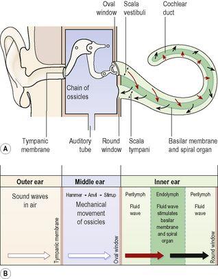
Figure 8.6
Passage of sound waves: A.
The ear with cochlea uncoiled.
B.
Summary of transmission.
The oval window is occluded by part of a small bone called the
stapes
and the round window, by a fine sheet of fibrous tissue.
Air reaches the cavity through the
pharyngotympanic
(
auditory
or
Eustachian
)
tube
, which extends from the nasopharynx. It is about 4 cm long and is lined with ciliated columnar epithelium. The presence of air at atmospheric pressure on both sides of the tympanic membrane is maintained by the pharyngotympanic tube and enables the membrane to vibrate when sound waves strike it. The pharyngotympanic tube is normally closed but when there is unequal pressure across the tympanic membrane, e.g. at high altitude, it is opened by swallowing or yawning and the ears ‘pop’, equalising the pressure again.
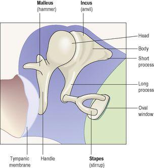
Auditory ossicles (
Fig. 8.3
)
These are three very small bones only a few millimetres in size that extend across the middle ear from the tympanic membrane to the oval window (
Fig. 8.1
). They form a series of movable joints with each other and with the medial wall of the cavity at the oval window. They are named according to their shapes.


