Ross & Wilson Anatomy and Physiology in Health and Illness (86 page)
Read Ross & Wilson Anatomy and Physiology in Health and Illness Online
Authors: Anne Waugh,Allison Grant
Tags: #Medical, #Nursing, #General, #Anatomy

Figure 8.3
The auditory ossicles.
The malleus
This is the lateral hammer-shaped bone. The handle is in contact with the tympanic membrane and the head forms a movable joint with the incus.
The incus
This is the middle anvil-shaped bone. Its body articulates with the malleus, the long process with the stapes, and it is stabilised by the short process, fixed by fibrous tissue to the posterior wall of the tympanic cavity.
The stapes
This is the medial stirrup-shaped bone. Its head articulates with the incus and its footplate fits into the oval window.
The three ossicles are held in position by fine ligaments.
Inner ear (
Fig. 8.4
)
The inner (internal) ear or labyrinth (meaning ‘maze’) contains the organs of hearing and balance. It is described in two parts, the
bony labyrinth
and the
membranous labyrinth
.
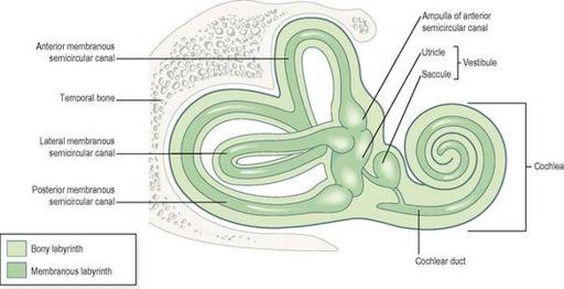
Figure 8.4
The inner ear
. The membranous labyrinth within the bony labyrinth.
Bony labyrinth
This is a cavity within the temporal bone lined with periosteum. It is larger than, and encloses, the membranous labyrinth of the same shape that fits into it, like a tube within a tube. Between the bony and membranous labyrinth there is a layer of watery fluid called
perilymph
and within the membranous labyrinth there is a similarly watery fluid,
endolymph
.
The bony labyrinth consists of:
•
the vestibule
•
the cochlea
•
three semicircular canals.
The vestibule
This is the expanded part nearest the middle ear. The oval and round windows are located in its lateral wall. It contains two membranous sacs, the utricle and the saccule, which are important in balance.
The cochlea
This resembles a snail’s shell. It has a broad base where it is continuous with the vestibule and a narrow apex, and it spirals round a central bony column.
The semicircular canals
These are three tubes arranged so that one is situated in each of the three planes of space. They are continuous with the vestibule.
Membranous labyrinth
This is a network of delicate tubes, filled with endolymph. It comprises:
•
the vestibule, which contains the
utricle
and
saccule
•
the cochlea
•
three semicircular canals.
The cochlea
A cross-section of the cochlea (
Fig. 8.5
) contains three compartments:
•
the scala vestibuli
•
the scala media, or
cochlear duct
•
the scala tympani.
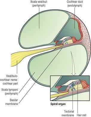
Figure 8.5
A cross-section of the cochlea showing the spiral organ (of Corti).
In cross-section the bony cochlea has two compartments containing perilymph: the scala vestibuli, which originates at the oval window, and the scala tympani, which ends at the round window. The two compartments are continuous with each other and
Figure 8.6
shows the relationship between these structures. The cochlear duct is part of the membranous labyrinth and is triangular in shape. On the
basilar membrane
, or base of the triangle, are
supporting cells
and specialised
cochlear hair cells
containing auditory receptors. These cells form the
spiral organ
(of Corti), the sensory organ that responds to vibration by initiating nerve impulses that are then perceived as hearing within the brain. The
auditory receptors
are dendrites of efferent (sensory) nerves that combine forming the cochlear (auditory) part of the vestibulocochlear nerve (8th cranial nerve), which passes through a foramen in the temporal bone to reach the hearing area in the temporal lobe of the cerebrum (see
Fig. 7.21, p. 150
).
Physiology of hearing
Every sound produces sound waves or vibrations in the air, which travel at about 332 metres per second. The auricle, because of its shape, collects and concentrates the waves and directs them along the auditory canal causing the tympanic membrane to vibrate. Tympanic membrane vibrations are transmitted and amplified through the middle ear by movement of the ossicles (
Fig. 8.6
). At their medial end the footplate of the stapes rocks to and fro in the oval window, setting up fluid waves in the perilymph of the scala vestibuli. Some of the force of these waves is transmitted along the length of the scala vestibuli and scala tympani, but most of the pressure is transmitted into the cochlear duct. This causes a corresponding wave motion in the endolymph, resulting in vibration of the basilar membrane and stimulation of the auditory receptors in the hair cells of the spiral organ. The nerve impulses generated pass to the brain in the cochlear (auditory) portion of the vestibulocochlear nerve (8th cranial nerve). The fluid wave is finally expended into the middle ear by vibration of the membrane of the round window. The vestibulocochlear nerve transmits the impulses to the auditory nuclei in the medulla, where they synapse before they are conducted to the auditory area in the temporal lobe of the cerebrum (see
Fig. 7.21, p. 150
). Because some fibres cross over in the medulla and others remain on the same side, the left and right auditory areas of the cerebrum receive impulses from both ears.
Sound waves have the properties of
pitch
and
volume
, or intensity (
Fig. 8.7
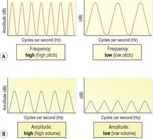
). Pitch is determined by the frequency of the sound waves and is measured in Hertz (Hz). Sounds of different frequencies stimulate the basilar membrane (
Fig. 8.6A
) at different places along its length, allowing discrimination of pitch.
Figure 8.7
Behaviour of sound waves. A.
Difference in frequency but of the same amplitude.
B.
Difference in amplitude but of the same frequency.
The volume depends on the magnitude of the sound waves and is measured in decibels (dB). The greater the amplitude of the wave created in the endolymph, the greater is the stimulation of the auditory receptors in the hair cells in the spiral organ, enabling perception of volume. Long-term exposure to very loud noise causes hearing loss because it damages the sensitive hair cells of the spiral organ.
Balance and the ear
Learning outcome
After studying this section, you should be able to:
describe the physiology of balance.
The semicircular canals and vestibule (
Fig. 8.4
)


