Ross & Wilson Anatomy and Physiology in Health and Illness (90 page)
Read Ross & Wilson Anatomy and Physiology in Health and Illness Online
Authors: Anne Waugh,Allison Grant
Tags: #Medical, #Nursing, #General, #Anatomy

Dark adaptation
When exposed to bright light, the rhodopsin within the sensitive rods is completely degraded. This does not affect vision in good light, when there is enough light to activate the cones. However, if the individual moves into a darkened area where the light intensity is insufficient to stimulate the cones, temporary visual impairment results whilst the rhodopsin is being regenerated within the rods, ‘dark adaptation’. When regeneration of rhodopsin has occurred, normal sight returns.
It is easier to see a dim star in the sky at night if the head is turned slightly away from it because light of low intensity is then focused on an area of the retina where there is a greater concentration of rods. If looked at directly the light intensity of a dim star is not sufficient to stimulate the less sensitive cones in the area of the macula lutea. In dim evening light, different colours cannot be distinguished because the light intensity is insufficient to stimulate colour-sensitive pigments in cones.
Breakdown and regeneration of the visual pigments in cones is similar to that of rods.
Binocular vision
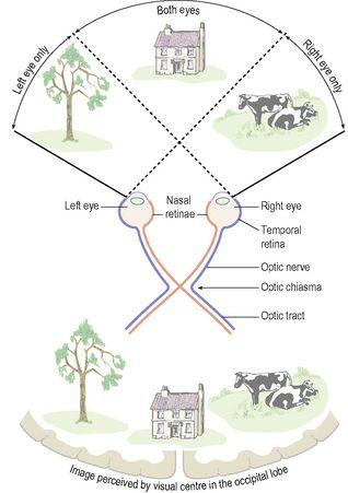
Binocular or stereoscopic vision enables three-dimensional views although each eye ‘sees’ a scene from a slightly different angle (
Fig. 8.19
). The visual fields overlap in the middle but the left eye sees more on the left than can be seen by the other eye and vice versa. The images from the two eyes are fused in the cerebrum so that only one image is perceived.
Figure 8.19
Parts of the visual field
– monocular and binocular.
Binocular vision provides a much more accurate assessment of one object relative to another, e.g. its distance, depth, height and width. People with monocular vision may find it difficult, for example, to judge the speed and distance of an approaching vehicle.
Extraocular muscles of the eye
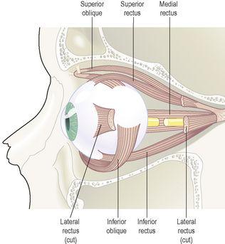
These include the muscles of the eyelids and those that move the eyeballs. The eyeball is moved by six
extrinsic muscles
, attached at one end to the eyeball and at the other to the walls of the orbital cavity. There are four
straight
(rectus) muscles and two
oblique
muscles (
Fig. 8.20
).
Figure 8.20
The extrinsic muscles of the eye.
Moving the eyes to look in a particular direction is under voluntary control, but coordination of movement, needed for convergence and accommodation to near or distant vision, is under autonomic (involuntary) control. Movements of the eyes resulting from the action of these muscles are shown in
Table 8.1
.
Table 8.1
Extrinsic muscles of the eye: their actions and cranial nerve supply
| Name | Action | Cranial nerve supply |
|---|---|---|
| Medial rectus | Rotates eyeball inwards | Oculomotor nerve (3rd cranial nerve) |
| Lateral rectus | Rotates eyeball outwards | Abducent nerve (6th cranial nerve) |
| Superior rectus | Rotates eyeball upwards | Oculomotor nerve (3rd cranial nerve) |
| Inferior rectus | Rotates eyeball downwards | Oculomotor nerve (3rd cranial nerve) |
| Superior oblique | Rotates eyeball downwards and outwards | Trochlear nerve (4th cranial nerve) |
| Inferior oblique | Rotates eyeball upwards and outwards | Oculomotor nerve (3rd cranial nerve) |
Nerve supply to the muscles of the eye
Nerves shown in
Table 8.1
supply the extrinsic muscles. The
oculomotor nerves
supply the
intrinsic eye muscles
of the iris and ciliary body.
Accessory organs of the eye
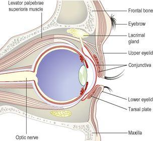
The eye is a delicate organ which is protected by several structures (
Fig. 8.21
):
•
eyebrows
•
eyelids and eyelashes
•
lacrimal apparatus.
Figure 8.21
Section of the eye and its accessory structures.
Eyebrows
These are two arched ridges of the supraorbital margins of the frontal bone. Numerous hairs (eyebrows) project obliquely from the surface of the skin. They protect the eyeball from sweat, dust and other foreign bodies.
Eyelids (palpebrae)
The eyelids are two movable folds of tissue situated above and below the front of each eye. On their free edges there are short curved hairs, the
eyelashes
. The layers of tissue forming the eyelids are:
•
a thin covering of skin
•
a thin sheet of subcutaneous connective (loose areolar) tissue
•
two muscles – the
orbicularis oculi
and
levator palpebrae superioris
•
a thin sheet of dense connective tissue, the
tarsal plate
, larger in the upper than in the lower eyelid, which supports the other structures
•
a lining of
conjunctiva
.
Conjunctiva
This is a fine transparent membrane that lines the eyelids and the front of the eyeball (
Fig. 8.21
). Where it lines the eyelids it consists of highly vascular columnar epithelium. Corneal conjunctiva consists of avascular stratified epithelium, i.e. epithelium without blood vessels. When the eyelids are closed the conjunctiva becomes a closed sac. It protects the delicate cornea and the front of the eye. When eyedrops are administered they are placed in the lower conjunctival sac. The medial and lateral angles of the eye where the upper and lower lids come together are called respectively the
medial canthus
and the
lateral canthus
.
Eyelid margins
Along the edges of the lids are numerous
sebaceous glands
, some with ducts opening into the hair follicles of the eyelashes and some on to the eyelid margins between the hairs.
Tarsal glands
(Meibomian glands) are modified sebaceous glands embedded in the tarsal plates with ducts that open on to the inside of the free margins of the eyelids. They secrete an oily material, spread over the conjunctiva by blinking, which delays evaporation of tears.
Functions
The eyelids and eyelashes protect the eye from injury:
•
reflex closure of the lids occurs when the conjunctiva or eyelashes are touched, when an object comes close to the eye or when a bright light shines into the eye – this is called the
corneal reflex
•
blinking at about 3- to 7-second intervals spreads tears and oily secretions over the cornea, preventing drying.
When the orbicularis oculi contract, the eyes close. When the levator palpebrae contract, the eyelids open (see
Fig. 16.58, p. 414
).
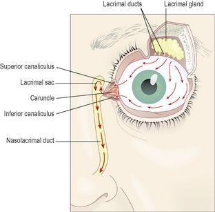
Lacrimal apparatus (
Fig. 8.22
)
For each eye this consists of:
•
1 lacrimal gland and its ducts
•
2 lacrimal canaliculi
•
1 lacrimal sac
•
1 nasolacrimal duct.
Figure 8.22
The lacrimal apparatus.
Arrows show the direction of the flow of tears.
The lacrimal glands
are exocrine glands situated in recesses in the frontal bones on the lateral aspect of each eye just behind the supraorbital margin. Each gland is approximately the size and shape of an almond, and is composed of secretory epithelial cells. The glands secrete
tears
composed of water, mineral salts, antibodies (immunoglobulins, see
Ch. 15
), and
lysozyme
, a bactericidal enzyme.
The tears leave the lacrimal gland by several small ducts and pass over the front of the eye under the lids towards the medial canthus where they drain into the
two lacrimal canaliculi
; the opening of each is called the
punctum
. The two canaliculi lie one above the other, separated by a small red body, the
caruncle
. The tears then drain into the
lacrimal sac
, which is the upper expanded end of the
nasolacrimal duct
. This is a membranous canal approximately 2 cm long, extending from the lower part of the lacrimal sac to the nasal cavity, opening at the level of the inferior concha. Normally the rate of secretion of tears keeps pace with the rate of drainage. When a foreign body or other irritant enters the eye the secretion of tears is greatly increased and the conjunctival blood vessels dilate. Secretion of tears is also increased in emotional states, e.g. crying, laughing.


