Fundamentals of Midwifery: A Textbook for Students (87 page)
Read Fundamentals of Midwifery: A Textbook for Students Online
Authors: Louise Lewis

BOOK: Fundamentals of Midwifery: A Textbook for Students
2.92Mb size Format: txt, pdf, ePub
phenylketonuria (PKU)
congenital hypothyroidism (CHT)
sickle cell disease (SCD)
cystic fibrosis (CF)
medium-chain acyl-CoA dehydrogenase deficiency (MCADD).
Throughout the UK the blood spot test is used to screen for PKU, cystic fibrosis and hypothy- roidism with some areas adding tests for MCADD, unusual haemoglobins (e.g. sickle cell) and galactosaemia (Wylie 2010). The purpose of the universal screening by this means is to detect disease as early as possible and therefore gain early referral and treatment which improves the outcome for the affected baby. Informed parental consent is essential and the blood sample must be taken safely and according to the protocol or there will be a requirement to repeat the sample causing both baby and parents unnecessary distress. The UK Newborn Screening Centre (2012) has published clear and detailed guidelines for professionals and information for parents and families (Activity 9.1).

Further reading activity
Read the Newborn Bloodspot Guidelines [Available online] http://newbornbloodspot.screening
.nhs.uk/bloodspotsampling

Figure 9.8
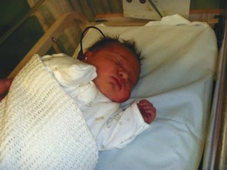
Baby receiving a hearing screen in the hospital, note the ear piece.

203
Newborn hearing screening
It is estimated that 1–3 of every 1000 newborn babies will be affected by congenital hearing loss which can be caused by neurological or conductive disorder. The hearing loss is mostly genetic in origin, but can also be caused by intrauterine infection (particularly cytomegalovirus) or prematurity. The universal screening programme has been in place in the UK since 2005, as early detection of deafness reduces its impact on language development. The test is generally carried out before the baby leaves hospital by trained personnel but can also be carried out in the home by health visitors and involves a small earpiece being inserted into the baby’s ear (see Figure 9.8). Two painless screening tests are carried out – the otoacoustic emissions test and the automated auditory brainstem response test – These will diagnose moderate to profound hearing loss.

Further reading activity
Read about the NHS Newborn Hearing Screening Programme [Available online] http://hearing
.screening.nhs.uk/
Advice for parents
Parents rely on healthcare professionals, particularly midwives, to provide them with relevant
information to enable them to keep their babies as safe as possible. As with all midwifery care this has to be tailored to individual needs. It should always include the information listed in Box 9.3, but questioning parents about issues that may concern them is also good practice. It is important to make sure that they feel they can discuss their worries without feeling silly as many parents feel that they should know how to care for their baby but in reality they have little knowledge and experience of small babies.

204

Box 9.3 Advice for parents
Cot death prevention information particularly in relation to positioning and co-sleeping (see Chapter 8 for more details)
Car safety
Appropriate room temperature and clothing guidance
Sterilising equipment (even if breastfeeding as breast pumps and bottles for expressed breastmilk may be used)
How to contact appropriate health care professionals in the event of any concerns about the health of the baby
All the advice provided to parents must be based on current evidence and where necessary midwives should utilise other members of the multidisciplinary team to provide information outside their own knowledge and expertise. A range of printed material is available and should be used to support the information given where appropriate; it is essential this is provided in appropriate format e.g. large print and other languages if necessary.
Detailed neonatal examination by the midwife
The more comprehensive examination advised by the National Screening Committee (NSC)
(2008; Public Health England 2013) has traditionally been undertaken by a paediatrician, but has now been incorporated as an extended role of the midwife who has undertaken further training to qualify for this. Some of the reasons reported why a midwife was preferred were: they impart more information whilst performing the examination; they spend more time at it; and they are often someone the mother knows. Part of the preparation for the examination should be gaining a good history, including a review of the maternal medical notes to highlight any risk factors that may need to be discussed with the parents (Tappero and Honeyfield 2003). It is here that a midwife undertaking the examination may also have an advantage over a paediatrician as they are likely to be already aware of the history from antenatal and intrapartum care.
The recommendation is for this full examination to be performed within 24 hours of birth, but some consideration should be given to the timing of the procedure. NICE 2006 guidelines recommend the full examination is done within 72 hours of birth and is repeated at the end of the postnatal period. As this examination has a greater chance of detecting a problem, it is of benefit to have both parents present or some alternative support for the mother. The midwife who is ward-based is more likely to be able to alter her schedule to enable this rather than a paediatrician with other time constraints and neonatal unit commitments.
On introduction to the parents, an explanation of and consent for the examination must be undertaken. Informed consent is a requirement for any procedure undertaken by a midwife (NMC, 2008) and for this consent to be valid, a number of facts must be made clear to the parents. It is important that parents are made aware that it is a midwife with further training who is undertaking the examination. Baston and Durward (2010) highlight that if parents are expecting a procedure to be performed by a doctor and are not made aware that it is not, this will render the consent invalid. Where it is an option, they should be offered the opportunity to have a paediatrician perform the examination if preferred. It should be explained that this is a screening test and as such may detect a problem. It has been suggested that similar to the ultrasound scans that are undertaken in early pregnancy, the parents are often not prepared
for the fact an abnormality may be detected (Baston and Durward 2010). This information should be given along with an explanation that if any concerns are highlighted they will be given further opportunity to discuss the problems and ask questions with a paediatrician. It is important to inform the parents that this examination may not detect some conditions which do not become apparent until later and they will be offered a further examination at 6 to 8 weeks (Davis and Elliman 2008). It should also be explained that if the examination is being performed after only six hours, there may be some anomalies detected that will not be found on a subsequent examination, such as a lax hip joint or soft heart murmur. It is useful to detail the four areas which will be examined in more detail than the initial examination: eyes, heart, hips and testes in boys with a brief explanation of the conditions being screened for. This intro- duction period also presents a useful time for more history gathering as the parents can be asked about any family history or observation of their new baby that has given them any cause for concern. Another good reason for waiting until both parents are present is that during this time observation of the parents’ facial features may explain what could otherwise have been considered anomalies in the infant (Baston and Durward 2010).
Communication appeared to be the key area where the midwives performing this examina- tion were appreciated more than the doctors (Murray et al. 2006). Having to break bad news to new parents is a very difficult thing to do and Robb (1999) suggests that parents do not always remember the exact words used to inform them of their baby’s disability, but they do remember the general approach and the staff’s attitude so this is a vital time and there is only one chance at it.
Other books
Sex on the Beach (Cosmo Red-Hot Reads from Harlequin) by Delphine Dryden
New tricks by Sherwood, Kate
The Bounty Hunter: Reckoning by Joseph Anderson
Crossing to Safety by Wallace Stegner
William Shakespeare's The Phantom Menace by Ian Doescher
Already Dead: A California Gothic by Denis Johnson
Breaking an Empire by James Tallett
Carolyn Davidson by The Forever Man
Believe It or Not by Tawna Fenske
Seeing Stars by Vanessa Grant