Secondary Schizophrenia (25 page)
Read Secondary Schizophrenia Online
Authors: Perminder S. Sachdev

also come to buttress this earliest of the functional
Hypofrontality
neuroimaging findings of schizophrenia research: use
of the functional near-infrared spectroscopy (fNIRS),
Starting with the initial observation using the
still in its nascency, has already provided support
133Xenon inhalation method by Ingvar and Franzen
for both the resting
[27]
and task-induced
[28, 29]
[4],
and the first independent confirmation using
hypofrontality and for the association of its sever-the newly available positron emission tomogra-
ity with the duration of illness. As a matter of fact,
phy (PET)
[5],
resting hypofrontality, or reduced
fNIRS studies in schizophrenia have thus far concen-frontal-to-occipital metabolic ratios, in patients with
trated mainly on the prefrontal cortex
[30, 31, 32,
schizophrenia has been reported in numerous studies
33]
, and the findings have been congruent with the
by various methodologies
[6, 7, 8, 9, 10].
Initially
extensive body of previously available neuroimaging
there were negative studies as well (e.g.
[11, 12, 13,
research.
14]).
Similarly, task-related hypofrontality under
various frontal lobe cognitive tasks in patients with
Cerebral disconnectivity and
schizophrenia has had almost as many proponents
[6, 9, 15, 16, 17, 18]
as detractors
[19, 20].
Reviews of
schizophrenia
progress in functional neuroimaging concluded that
Schizophrenia, since its very nosological inception,
hypofrontality had been the most consistent finding in
had generally been considered a grey matter disease.
schizophrenia
[21, 22];
yet another contemporaneous
Despite decades of intense neuropathological inves-review estimated that hypofrontality was found in
tigations that failed to establish anatomical basis of
only about one-third of published reports
[23].
the illness, the vast majority of neuroimaging stud-A more stringent meta-analysis of activation paties for a long time concentrated almost exclusively on
terns in 103 suitable voxel-based 133Xenon inhala-the grey matter. Initial limitations of the available neu-tion, single photon emission computed tomography
roimaging methodologies did confine their applica-
(SPECT), and PET (15O2 and FDG) studies of pre-
tion mainly to the analysis of the ventricular system
frontal activation supported both resting and task-and anatomically bounded grey matter. Things began
activated hypofrontality in patients with schizophre-to change only with the advent of the newer tomonia
[24].
Moreover, a positive association of resting
graphic techniques for functional neuroimaging –
hypofrontality with duration of illness was also sug-based on the positron-emission scintigraphy and later
gested in this analysis and may provide an explana-on the ultrafast sequences of magnetic resonance sig-tion for some of the earlier discrepancies. Whether
nal (MRS) acquisition. These methodological devel-this longitudinal pattern reflects the actual chronicity
opments were paralleled by a shift of overall empha-
61
of the illness or disease-independent treatment effects
sis from localization of cerebral function to hodology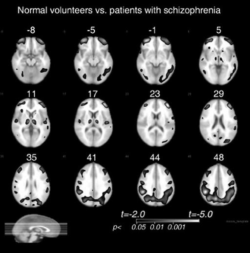
The Neurology of Schizophrenia – Section 2
Figure 5.2
Group comparison of
patients with schizophrenia and normal
controls. Areas where patients (n
=
59)
are significantly lower than normals (n
=
70) are shown with a black edge and light
interior corresponding to the t grey-scale
bar in the lower right. The Talairach z level
is shown in mm above each slice image.
The background to the patches identified
as statistically significant is the Montreal
Neurological Institute anatomical MRI
brain.
in neuroanatomy as a scientific discipline
[34].
The
stemming from the disturbed neuronal migration in
newly popular hodological, or connectionist, approach
the second trimester, may be at play
[44, 45, 46, 47].
to functional neuroanatomy naturally led to refreshed
Indeed, supportive of the latter hypothesis, experimen-interest in the so-called disconnection syndromes
[35]
tal miswiring of prefrontal efferents in Mongolian ger-and it did not take long for schizophrenia to be pos-bils was recently induced by methamphetamine intox-tulated as one
[36, 37, 38, 39].
In fact, ideas regard-ication in the early postnatal period
[6].
Still oth-ing interhemispheric disconnectivity in schizophrenia
ers point to the possibility that dysmyelination may
had circulated even before the rise of modern neu-be pivotal in the pathophysiology and even etiol-roimaging techniques
[40].
ogy of the illness
[48]
, or – in other words – that
One of the most consistent theoreticians to regard
schizophrenia is a white-matter disease par excellence.
schizophrenia as a disconnection syndrome – Karl
This view draws its supportive evidence from a host
Friston – has envisioned it as a disorder of functional
of recent cerebral gene expression, diffusion-weighted,
integration, the central pathophysiological mechanism
and magnetization transfer imaging studies
[49].
This
being that of disturbed synaptic plasticity
[37, 41, 42,
is further bolstered by a very recent discovery that
43]
. Friston regards schizophrenia as a primary dis-unlike the many grey-matter findings, relative glucose
order of synaptic transmission resulting in discon-metabolism in patients with schizophrenia appears
nectivity. He emphasizes dysfunctional regional inte-to be increased in the white matter
[50].
Irrespec-gration as opposed to dysfunctional regional special-tive of the proposed pathophysiology, the disconnec-ization (which he considers to be a secondary feation hypothesis has served to shift the scientific inter-ture), and functional as opposed to anatomical dis-est away from structural volumetrics to functional
connectivity. Some other authors suggest that devel-interregional interactions. It also helps bridge the gap
62
opmental anatomical disconnectivity (i.e. miswiring),
between the so-called “functional” and “organic” views
Chapter 5 – Functional neuroimaging in schizophrenia
of schizophrenia as this distinction becomes less clear
ing multivariate analytic techniques to the hypothesis-when disconnection is considered to be the pathophys-independent voxel-to-voxel measurements with the
iological basis.
creation of synchronization maps.
The neuroimaging methods for studying regional
Finally, evaluation of interregional correlations
interconnectivity include evaluation of individual
in volumetric data, derived from structural MRI
regions of interest and use of bivariate correlation coef-analyses, may also be viewed as providing task-
ficients or higher-order factor analysis, path analysis,
independent information on sustained, tonic activa-and other multivariate techniques to infer the interre-tion of functional networks, consistent enough to
gional relationships among them. Thus, interregional
result in correlated trophic influences and thus point
correlation matrices of glucose metabolic rates have
to abnormalities of sustained networking in patients
been widely used and validated in the assessment of
with schizophrenia relative to healthy controls. The-functional neural systems
[51, 52, 53, 54, 55,
56].
Pair-oretically, this approach allows for evaluation of
wise interregional metabolic correlations are thought
chronically engaged, abnormal cerebral networks
to reflect functional connectivity, but these do not
independently of a task. In our study comparing vol-allow any inferences on the directionality of the func-umetric and metabolic thalamocortical intercorrelational regional interactions. Structural equation mod-tions in healthy subjects during a ubiquitous verbal
eling based on a priori neuroanatomical assumptions
learning task, metabolic intercorrelations were much
has been employed in order to account for the direc-more widespread and numerous in comparison to the
tionality and strength of the interregional influences,
correlations of regional volumes
[66].
The idea that
that is, the so-called effective connectivity
[57, 58, 59,
grey-matter volumetric abnormalities may be medi-
60].
These methods, initially developed for PET, have
ated by consistent functional activations is prelim-later been applied to time-series data derived from the
inarily supported by a recent study reporting that
functional MRI, and, rather than relying on manu-an association of thought disorder in schizophren-ally applied regions of interest, now primarily exploit
ics with grey-matter reductions in planum tempo-
the statistical parametric mapping (SPM) analysis of
rale was mediated by posterior temporal hyperacti-voxel-to-voxel correlations at rest and under imposed
vation in BOLD signal
[67].
In group comparisons,
cognitive tasks
[61, 62, 63].
significant direct interregional correlations present
Use of continuous cognitive tasks allows for detec-only in patients with schizophrenia would signify
tion of relative activation failures at locations normally
abnormal reliance on alternative strategies for com-activated by a task or aberrant regional recruitment
pensatory information processing. The absence of nor-that may be conceptualized as pathological reliance
mal interregional correlations in patients would sig-on alternative networks in a compensatory effort. The
nify reduced interregional connectivity
[68, 69].
Fol-major difficulty with this approach lies in the compar-lowing is the review of selected experimental literature
ison of groups of subjects differing in test performance
on the aforementioned aspects of functional regional
[64],
whereby the differences in performance mediate
integration in schizophrenia.
the differential patterns of activation
[65].
In addition,
association of these pathologically recruited networks
Aberrant regional recruitment
with specific clinical symptoms (e.g., hallucinations or
under a task
specific delusional beliefs) or cognitive deficits (e.g., in
short-term memory, implicit learning, or visual recognition) may allow for certain anatomical localization
PET and SPECT
of the clinical semiotics.
Countless assessments of regional changes in glu-Another approach to studying interregional inte-
cose metabolism or oxygen utilization have docu-
gration in the brain is EEG evaluation of task-
mented aberrant regional activations in patients with
induced temporal synchronization of electrical activ-schizophrenia in association with a vast array of cog-ity across bandwidths and regions of interest. As
nitive tasks and within a multitude of surveyed cere-with other functional neuroimaging methods, this
bral structures
[70, 71].
These include the frontal lobe,
may be accomplished by using bivariate correlative
mainly subregional prefrontal cortex
[72, 73],
anterior
or phase coherence measures of time series data col-cingulate gyrus
[74, 75, 76],
temporal lobe
[77],
pari-
63
lected from preselected pairs of electrodes or by apply-etal lobe, posterior cingulate gyrus
[75],
occipital lobe
The Neurology of Schizophrenia – Section 2
[77],
striatum
[78],
cerebellum
[79]
, thalamus and its
Resting disconnectivity