Secondary Schizophrenia (27 page)
Read Secondary Schizophrenia Online
Authors: Perminder S. Sachdev

Brodmann areas
[68, 69]
documented reduced asso-Reality distortion was associated with hyperac-
ciations among proximate regions within the frontal
tivation in the medial prefrontal cortex
[172]
and
and temporal lobes, and strengthening of more dis-a persecutory attributional stance in patients was
tant (interlobar) fronto-temporal, as well as fronto-accompanied by more severe prefronto-amygdalar
parietal and temporo-occipital intercorrelations, albeit
disconnection
[173].
less prominently, in patients with schizophrenia.
Impaired connectivity between the anterior-
Fronto-temporal grey matter volume dissociations in
cingulate and supplementary motor areas was related
schizophrenics have been reported in some other
to negative symptoms. The poor functional outcome
investigations
[44,
152, 153].
in schizophrenia overall was associated with more
Thalamocortical dissociations in patients with
severe hypoactivation in the temporal lobe and cingu-schizophrenia, assessed in another two studies from
late gyrus, as well as more significant hypofrontality at
our laboratory
[154, 155],
were found to be rather
rest, as compared to the good-outcome patient group
widespread, especially with the prefrontal, medial-
[174]
. Finally, one of the often-reported schizophrenia
temporal, and cingulate-cortical regions in both
endophenotypes – impaired inhibition of saccadic
hemispheres and, for the pulvinar with the occip-eye movements – was ascribed to failure to activate
ital and orbito-frontal cortices in the right hemi-the lentiform nuclei, thalami, and the left inferior
sphere. The latter pulvino-cortical dissociations were
frontal gyrus in response to an antisaccadic task
proposed to be pathogenetically related to visual
[175].
attentional deficits amply described in patients with
schizophrenia.
Receptor occupancy evaluation
Functional imaging correlates of
with PET
Dopamine D
clinical symptomatology
2 receptor binding in neuroleptic-naive
patients with schizophrenia has in recent years been
A number of functional neuroimaging studies have
studied using raclopride-11C as the ligand. Reduced
in recent years increasingly focused on relationships
ligand binding has been reported in the thalamus
[176,
between regional abnormalities and specific clinical
177, 178],
especially within the left mediodorsal and
symptoms or syndromes of schizophrenia
[156].
Audi-pulvinar nuclei
[176],
as well as the anterior cingulate
tory hallucinations have been consistently related to
[176, 179],
amygdala
[176]
, but not the caudate
[177].
activations in the left superior temporal gyrus and,
The biggest effect has thus far been reported in the
66

Chapter 5 – Functional neuroimaging in schizophrenia
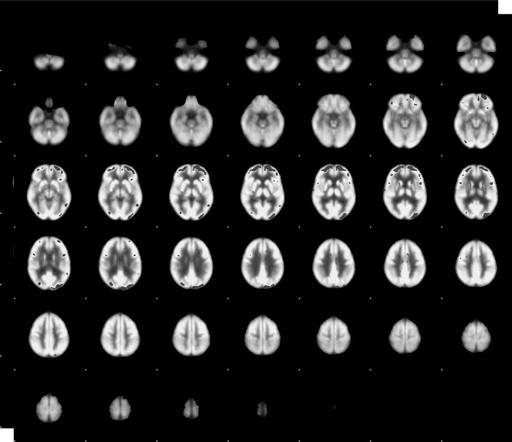
Figure 5.3
Significance probability mapping
[184]
test areas of decrease in FDG relative metabolic rate and D2 receptor binding. (See color
plate section.)
thalamus. One study that found no regional between-
Conclusion and new directions
group differences in the raclopride C11 binding
[180]
At the present time, all functional neuroimaging
did report a significant direct correlation between the
modalities are in transition from being strictly
ligand binding in the frontal lobe and the positive
research tools to assisting clinical diagnosis and
symptom severity in schizophrenics.
treatment choice. Work is being done on developing
Preliminary investigations with some other lig-
fMRI classificatory instruments aimed at image-based
ands have been less conclusive, with reports of either
identification of patients with schizophrenia
[185]
no intergroup differences
[181]
or elevated binding
and on PET prediction of response to neuroleptic
[182]
for serotonin 1A receptors, as well as the as-yet
treatment
[186].
This may be reasonably expected to
unreplicated finding of decreased frontal lobe binding
eventually make functional neuroimaging clinically
for histamine H1 receptors
[183].
useful for diagnosis and treatment.
67
The Neurology of Schizophrenia – Section 2
References
9. Ortu˜no F., Moreno-I˜niguez M.,
sorting activated regional cerebral
Millan M.,
et al.
Cortical blood
blood flow in first break and
1. Gloor, P. Hans Berger and the
flow during rest and Wisconsin
chronic schizophrenic patients
discovery of the electro-
Card Sorting Test performance in
and normal controls. Schizophr
encephalogram.
schizophrenia. Wien Med
Res, 1996.
19
:177–87.
Electroencephalography and Clin
Wochenschr, 2006.
156
:179–84.
Neurophysiol, 1969. (Suppl)
18. Weinberger D. R., Berman K. F.,
28
:1–36.
10. Wolkin A., Jaeger J., Brodie J. D.,
Zec R. F. Physiological
et al.
Persistence of cerebral
dysfunction in dorsolateral
2. Wortis J., Bowman K. M.,
metabolic abnormalities in
prefrontal cortex in
Goldfarb W. Human brain
chronic schizophrenia as
schizophrenia. I. Regional
metabolism: normal values and
determined by positron emission
cerebral blood flow evidence. Arch
values in certain clinical states.
tomography. Am J Psychiatry,
Gen Psychiatry, 1986.
43
:
Am J Psychiatry, 1940–1.
97
:
1985.
142
:564–71.
114–24.
552–65.
3. Kety S. S., Woodford R.,
et al.
11. Ebmeier K. P., Lawrie S. M.,
19. Callicott J. H., Bretolino A.,
Cerebral blood flow and
Blackwood D. H. R.,
et al.
Mattay V. S.,
et al.
Physiological
metabolism in schizophrenia. Am
Hypofrontality revisited: a high
dysfunction of the dorsolateral
J Psychiatry, 1948.
104
:
resolution single photon emission
prefrontal cortex in schizophrenia
765–70.
computed tomography study in
revisited. Cereb Cortex, 2000.
schizophrenia. J Neurosurg
10
;1078–92.
4. Ingvar D. H., Franzen G.
Psychiatry, 1995.
58
:452–6.
Abnormalities of cerebral blood
20. Walter H., Wunderlich A. P.,
flow distribution in patients with
12. Gur R. E., Skolnick B. E., Gur R.
Blankenhorn M.,
et al.
No
chronic schizophrenia. Acta
C.,
et al.
Brain function in
hypofrontality, but absence of
Psychiatr Scand, 1974.
50
:
psychiatric disorders. I. Regional
prefrontal lateralization
425–62.
cerebral blood flow in medicated
comparing verbal and spatial
schizophrenics. Arch Gen
working memory in
5. Buchsbaum M. S., Ingvar D. H.,
Psychiatry, 1983.
40
:1250–54.
schizophrenia. Schizophr Res,
Kessler R.,
et al.
Cerebral
2003.
61
:175–84.
glucography with positron
13. Matthew R. J., Duncan G. C.,
emission tomography. Use in
Weinman M. L.,
et al.
Regional
21. Andreasen N. C., O’Leary D. S.,
normal subjects and in patients
cerebral blood flow in
Flaum M.,
et al.
Hypofrontality in
with schizophrenia. Arch Gen
schizophrenia. Arch Gen
schizophrenia: distributed
Psychiatry, 1982.
39
:251–59.
Psychiatry, 1982.
39
:1121–4.
dysfunctional circuits in
neuroleptic-na¨ıve patients.
6. Andreasen N. C., Rezai K., Alliger
14. Sheppard G., Gruzelier J.,
Lancet, 1997.
349
:1730–4.
R.,
et al.
Hypofrontality in
Manchanda R.,
et al.
15O positron
neuroleptic-naive patients and in
emission tomographic scanning
22. Velakoulis D., Pantelis C. What
patients with chronic
of predominantly never-treated
have we learned from functional
schizophrenia. Assessment with
acute schizophrenic patients.
imaging studies in schizophrenia?
xenon 133 single-photon emission
Lancet, 1983. 1448–52.
The role of frontal, striatal and
computed tomography and the
15. Buchsbaum M. S., Nuechterlein
temporal areas. Aust N Z J
Tower of London. Arch Gen
K. H., Haier R. J.,
et al.
Glucose
Psychiatry, 1996.
30
:195–209.
Psychiatry, 1992.
49
:943–58.
metabolic rate in normals and
23. Chua S. E., McKenna P. J.
7. Bagorda F., Teuchert-Noodt G.,
schizophrenics during the
Schizophrenia – a brain disease?
Lehmann K.. Isolation rearing or
continuous performance test
A critical review of structural and
methamphetamine traumatisation
assessed by positron emission
functional cerebral abnormality in
induce a “disconnection” of
tomography. Br J Psychiatry, 1990.
the disorder. Br J Psychiatry, 1996.
prefrontal efferents in gerbils:
156
:216–27.
166
:563–82.
implications for schizophrenia. J
16. Hazlett E. A., Buchsbaum M. S.,
24. Hill K., Mann L., Laws K. R.,
et al.
Neural Transm, 2006.
113
:365–79.
Jeu L. A.,
et al.
Hypofrontality in
Hypofrontality in schizophrenia: a
8. Farkas T., Wolf A. P., Jaeger J., et
unmedicated schizophrenia
meta-analysis of functional
al. Regional brain glucose
patients studied with PET during
imaging studies. Acta Psychiatr
metabolism in chronic
performance of a serial verbal
Scand, 2004.
110
:243–56.
schizophrenia. A positron
learning task. Schizophr Res, 2000.
25. Molina V., Sanz J., Reig S.,
et al.
emission transaxial tomographic
43
:33–46.
Hypofrontality in males with
study. Arch Gen Psychiatry, 1984.
17. Steinberg J. L., Devous Sr. M. D.,
first-episode psychosis. Br J
68
41
:293–300.
Paulman R. G. Wisconsin card
Psychiatry, 2005.
186
:203–8.
Chapter 5 – Functional neuroimaging in schizophrenia
26. Molina V., Sanz J., Sarramea F.,
35. Catani M., ffytche D. H. The rises
46. Jones, E. (1995) Cortical
et al.
Marked hypofrontality in