Wallach's Interpretation of Diagnostic Tests: Pathways to Arriving at a Clinical Diagnosis (525 page)
Authors: Mary A. Williamson Mt(ascp) Phd,L. Michael Snyder Md

The majority of CKD cases are due to glomerular disease, tubular or interstitial disease, or long-standing obstructive uropathy.
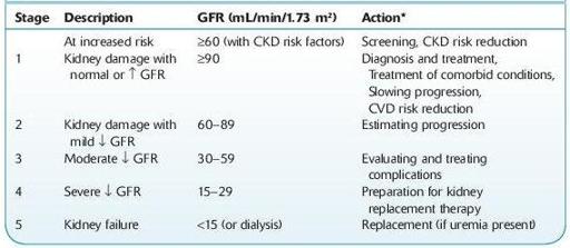
TABLE 12–3. Classification and Clinical Action Plan for Chronic Kidney Disease

Shaded area identifies patients who have chronic kidney disease; unshaded area designates individuals who are at increased risk for developing chronic kidney disease.
*Includes actions from preceding stages.
GFR, glomerular filtration rate; CKD, chronic kidney disease; CVD, cardiovascular disease.
Source: National Kidney Foundation. K/DOQI clinical practice guidelines for chronic kidney disease: evaluation, classification and stratification.
Am J Kidney Dis.
2002;39(Suppl 1):S1–S266.
Who Should Be Suspected?
Patients who have had acute renal injury or glomerulonephritis.
Patients with diseases that can initiate or propagate kidney disease, such as diabetes or hypertension.
Those in whom symptoms develop insidiously suggesting uremia (easy fatigability, anorexia, vomiting, mental status changes, seizures) or generalized edema.
Those in whom renal function or urinalysis abnormalities are discovered incidentally.
Those with a family history of CKD or have a congenital renal abnormality.
Laboratory Findings
Laboratory studies are indicated once renal disease is suspected. Until renal insufficiency is severe, adaptation of tubular function allows excretion of relatively normal amounts of water and sodium.
Serum creatinine and BUN increase in parallel.
Creatinine clearance, established via well-accepted formulas, is used to estimate the GFR (estimated GFR or eGFR). This parameter is generally considered the best index of overall kidney function and repeated determinations, in conjunction with creatinine measurement, establish whether the patient has stable or progressive disease. GFR has no etiologic diagnostic value.
Albuminuria is a marker of kidney damage that is commonly determined by measuring albumin/creatinine ratio (ACR) in untimed “spot” urine. An ACR cutoff of 30 mg/g indicates abnormality (see Figure
12-2
).